Download
1 / 40
790 likes | 2.44k Vues
Treatment principles of maxillofacial trauma. Reporter : Ho-Tai Wu. Evaluation of p’t with facial trauma Classification of facial fractures Treatment of facial fractures. Maxillofacial Injuries. Treatment divided into following phases Emergency or initial care Early care Definitive care.

E N D
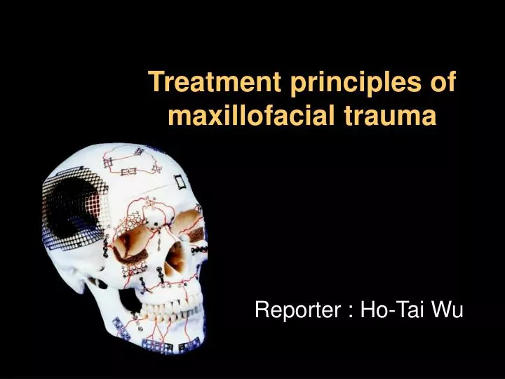
Treatment principles of maxillofacial trauma Reporter : Ho-Tai Wu
Evaluation of p’t with facial trauma • Classification of facial fractures • Treatment of facial fractures
Maxillofacial Injuries • Treatment divided into following phases • Emergency or initial care • Early care • Definitive care Trauma ER Stable OMFS,ENT, Plastic
Emergency Care • Preserve the airway • Control of hemorrhage • Prevent or control shock • C-Spine stabilization • Control of life-threatening injuries • head injuries, chest injuries, compound limb fractures, intra-abdominal bleeding
Emergency Care Preserve the airway • Existence & identification of obstruction • Manually clear of fractured teeth, blood clots, dentures • Endotracheal intubation & packing of oronasal airway
Emergency Care Control of hemorrhage • Extensive vascularity of head & neck may lead to massive blood loss • Monitor vital signs closely • Intravenous infusion • Penetrating injuries need to be explored • Arteriogram • Esophagram
Emergency Care Prevent or control shock • Hemorrhage most common cause of shock after injury • Multiple injury patients have hypovolemia • Goal is to restore organ perfusion
Emergency Care C-Spine stabilization • Avoid any movement of spinal column • Until rule out C-spine Fx • Lateral C-spine radiographs • CT of C-spine • Neurologic exam
Early Care • Emergency care has stabilized patient • Initial stabilization of fractures • Debridement & dressing of soft tissues • Elective tracheostomy • Physical exam & history • Laboratory tests • Complete head & neck examination • Diagnosis of maxillofacial injuries
History • Patient, witness or family member • How did the accident occur ? • When did the accident occur ? • What are the specifics of the injury ? • Was there a loss of consciousness ? • What symptoms are now ?
Physical examination • Inspection • Hemorrhage • Otorrhea • Rhinorrhea • Contour deformity • Ecchymosis • Edema • Continuity defects • Malocclusion
Physical examination • PALPATION • “Step” Defect • Crepitus • Bony segments • Subcutaneous emphysema • Mobility
Physical examination • Neurological examination • Visual or pupillary changes (CN II III IV VI) • Abnormalities of ocular movements (neurologic, orbital area fractures ) • Motor function of the facial muscle (CN VII) • Muscles of mastication (CN V) • Sensation the facial area (CN V)
Maxilla • Mobility of the maxilla either as an isolated zygoma or nasal bones • Palpated for step deformites in forehead, orbital rim, nasal or zygoma areas • Evaluation of the nose, nasal spetum and paranasal structures • Intercanthal distance (naso-orbital ethmoid injures) Mandible • TMJ • Occlusion plane • Laceration of oral cavity • Mobility and missing of the teeth
DIAGNOSTIC IMAGING Mandible fractures : PA view Lateral oblique view Towne’s view Panoramic view Occlusal view CT
DIAGNOSTIC IMAGING Midface fractures Water’s view Lateral skull view PA skull view Submental vertex view CT scans 3-D reconstruction
Evaluation of p’t with facial trauma • Classification of facial fractures • Treatment of facial fractures
Mandibular Fractures • 50% of mandibular fractures are multiple • Examine patient and radiographs closely and suspect additional fractures
Mandibular Fractures Condylar Angle Body Symphyseal Alveolar Ramus Coronoid precess
Mandibular Fractures Greenstick Simple Compound Comminuted
Mandibular Fractures Horizontally favorable Horizontally unfavorable Vertically favorable Vertically unfavorable
Mandibular Fractures • Radiographic evaluation
Midface Fractures • Three buttresses allow face to absorb force • Nasomaxillary (medial) buttress • Zymaticomaxillary (lateral) buttress • Pyterigomaxillary (posterior) buttress
LeFort I Transverse Maxillary Lefort II Pyramidal Lefort III Craniofacial Dysjunction Zygomatic Complex Naso-orbital/Ethmoid Midface Fractures Occlusion involved
Lefort I FractureTransverse Maxillary above the level of teeth Lefort II FracturePyramidal at level of nasal bones Lefort III FractureCraniofacial Dysjunction at orbital level (Rene’ Lefort, 1901)
Zygomatic Complex Naso-orbital/Ethmoid
Evaluation of p’t with facial trauma • Classification of facial fractures • Treatment of facial fractures
Goals of the treatments • Rapid bone healing • Return of normal appearance • Masticatory and nasal function • Restoration of speech • Acceptable esthetic
Basic surgical principle • Reduction of the fracture • Fixation of the bony segment • Stabilization of the bony segment • Immobilization of segments • Preoperative occlusion must be restored • Infection in the area must be eradicated
Basic surgical procedure 1. Place the teeth in the proper occlusion 2. Appropriated reduction of bony fractures 3. Bony repair should also precede soft tissue repair 4. Fractures treated as soon as the p’t condition permits 5. Rigid fixation begins in the area where fractures can e most easily stabilized .
Principle of mandibular fracture • Closed reduction 1. IMF ( Prefabricated arch bar ) 2. Ivy loop wiring 3. Continuous loop wiring 4. Heavy elastic traction (pull the bony segments) 5. Circummandibular wiring (edentulous p’t) 6. Lingual or occlusal splint (children)
Principle of mandibular fracture • Open reduction • Continued displacement of the bony segments • Unfavorable fracture (angle fracture) • Intraosseous wiring + IMF (3 to 8 weeks) • Rigid internal fixation these method use bone plates, bone screws or both to fix the fracture. • Proper occlusal established before reduction stabilization and fixation of the bony segment
Advantages of rigid fixation • IMF is eliminated or reduced • Improved postoperative nutrition • Improved postoperative hygiene External skeletal fixation • Appearance and inconvenience • Functional movement • Comminuted fracture
Principle of midface fracture • Zygomaticomaxillary buttress • Zygomaticofrontal area • Orbital rim area
Principle of midface fracture • Occlusion related (Le fort fractures) • Rehabilitation of occlusion relationship • Reduction of bone segment • Stablization of fractured segments
Principle of midface fracture Stablization of fractured segments • DirectIntraosseous wiring • Suspensionwiring technique • Bone plate & screws
Principle of midface fracture • Zygomatic Complex & Naso-orbital- Ethmoid fracture • Repair functions of eye, nose & mastication. • Acceptable esthetic • Prevent interfere to coronoid process (Zygomatic arch)
Conclusion Goals of the treatments • Rapid bone healing • Return of normal appearance • Masticatory and nasal function • Restoration of speech • Acceptable esthetic ANATOMY !!