Download
1 / 13
160 likes | 502 Vues
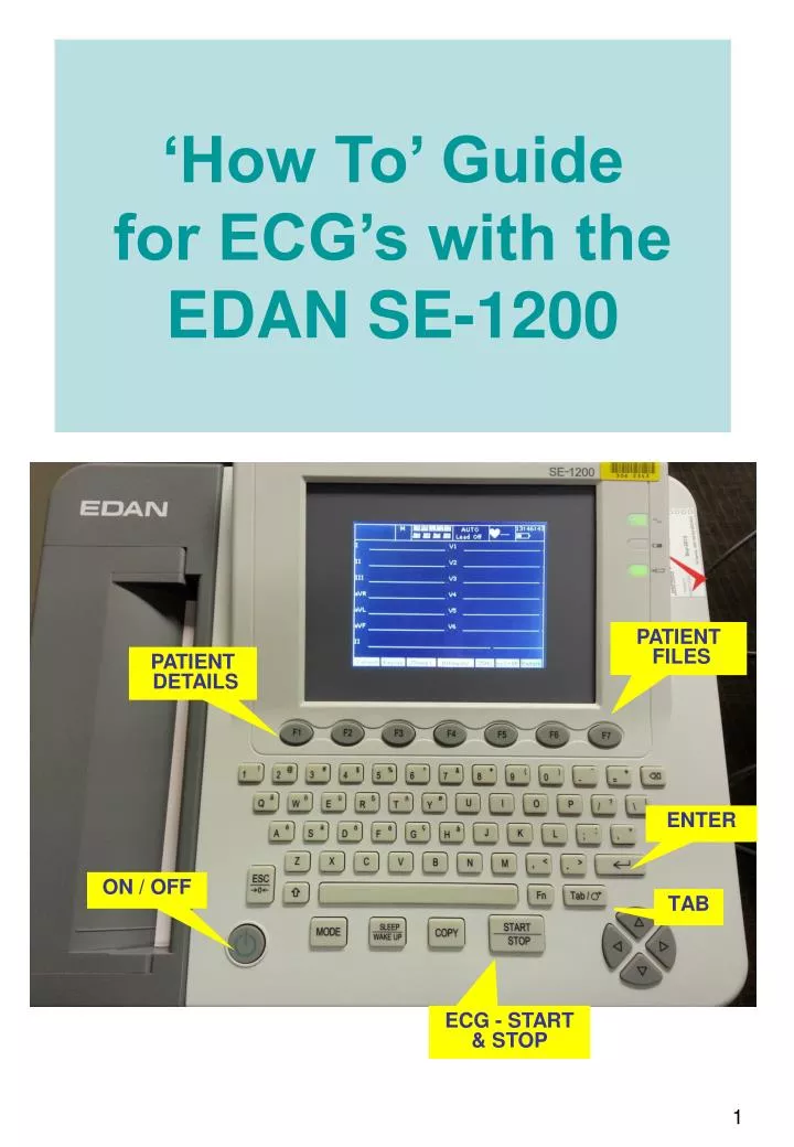
‘How To’ Guide for ECG’s with the EDAN SE-1200. TAB. PATIENT FILES. PATIENT DETAILS. ENTER. ON / OFF. ECG - START & STOP. PATIENT DATA PRESS F1 TO OPEN PATIENT DATA SCREEN. TAB ACROSS TO EACH FIELD. PATIENT ID. TO COMMENCE WITH HOSPITAL PREFIX. Exam room = Ward.

E N D
‘How To’ Guide for ECG’s with the EDAN SE-1200 TAB PATIENT FILES PATIENT DETAILS ENTER ON / OFF ECG - START & STOP
PATIENT DATAPRESS F1 TO OPEN PATIENT DATA SCREEN TAB ACROSS TO EACH FIELD PATIENT ID. TO COMMENCE WITH HOSPITAL PREFIX Exam room = Ward TAB TO OK THEN PRESS “ENTER Exam • Patient Details • To enter patient details, Tab across to each field. • If you tab past a field, either Shift + tab to move the cursor back or continue tabbing through all fields until returning to the missed one. • Once all fields have been completed, tab to OK then press enter button. • This will return screen to ECG Tracing
TELSTRA DEVICE Ensure Telstra Device is turned on Check that the device states connected Check signal strength indicated by number of bars in top left corner Check that signal tower has a small 1 beside tower Check for battery strength DO NOT turn off device. ENSURE ALL ANCILLARY DEVICES ARE CONNECTED ON / OFF Power MODEM Power Light will be on LAN light will be on WLAN light will be orange and changes to green when Telstra device is transmitting – returns to orange after
DOING THE ECG Prior Ensure all equipment on ECG trolley: ECG electrodes, Chlor-Hex swabs, clippers and blades; check paper level in machine Explain ECG procedure to patient Enter patient data into machine – all fields MUST be filled (ref p2) Wash hands Position patient in a supine position unless excessive dyspnoea is evident Expose the minimum amount of area required for the accurate positioning of the leads to ensure patient privacy and warmth Shave chest and limb hair as required – electrodes need good skin contact for good ECG tracing Important Be aware of any cardiac symptoms at the time of recording, such as chest pain, palpitations or shortness of breath Symptoms must be hand written on the printout and reported to nurse in charge Check Telstra Device connection – can be manually transmitted later Ensure good quality ECG trace before printing/sending. Remove electrodes: re-stick electrodes on plastic film from packet, place patient label on back of film and store in patient folder Cover the patient and return them to a position of comfort Wash Hands Return ECG trolley to storage. Plug In white power cord. Wipe leads with detergent wipes, replace equipment as required and leave trolley clean
ECG Placement Limb Leads Apply limb ECG electrodes to the soft tissue of limbs or outer torso LA: black– Left Arm LL: red – Left Leg RA: white – Right Arm RL: green – Right Leg Chest Leads V1: red – 4th Intercostal space, right sternal border. V2:yellow– 4th Intercostal space, left sternal border. V3: blue – Midway between V2 and V4. V4: green – 5th Intercostal space, midclavicular line. V5: orange – Anterior axillary line, horizontal to V4 V6: purple– Mid axillary line, horizontal to V4 and V5. (Clip chest hair if necessary to ensure good ECG tracing) Angle of Louis / Sternal Angle Start counting ICS from here
Chest Lead Placement –To position chest leads accurately it is important to identify the Angle of Louis Angle of Louis / Sternal angle The junction of the manubrium and the body of the sternum, it forms a ridge on the sternum, at the level of the second rib Find it on yourself: place fingers gently at the base of your throat (sternal notch) and move fingers downward over the top of the sternum until you feel a distinct bony ridge. Counting Intercostal Spaces (ICS) The 2nd rib is continuous with the sternal angle; hence the rib space above the sternal angle is the 1st ICS and below is the 2nd ICS. Continue counting down to the 4th ICS for V1 & V2, positioned at the edge of the sternum. The 5th ISC is the position of V4, in line with the middle of the clavicle. V3, V5 & V6 V3 sits midway between V2 & V4, diagonally. Continue in a horizontal line from V4. With the patient’s arm at their left side, follow the crease line in their armpit down the front of the their chest. This is the position of V5. Continue further in a horizontal line from V4 and in line with the centre point of the axilla. This is the position of V6, at the mid axillary line. Angle of Louis Start counting ICS from here
RECORDING THE ECG Check to ensure Modem and Telstra device are connected To record an ECG press the STOP / START button Appearing at the top of screen, under AUTO: Analysing Printing ECG Printout will occur automatically Transmitting Will be displayed until transmission complete If transmitting NOT successful, ECG is stored, accessed and manually transmitted via F7, Patient Files Under AUTO Analysing Printing Trans Will appear
Screen will change to bring up list of patients The following functions are at bottom of screen Trans All – this allows all listed ECG’s to be transmitted to Cardioscan. Export All – This allows all ECG’s to be exported to different file Del All – This allows all highlighted ECG’s to be deleted Select - This highlights and selects the patient required Search – this allows you to look for a patient by either Name, ID , or Time. Import – This allows data to be imported from external source Return – this allows a return to previous page. If a patient ECG has not transmitted successfully, there will be a blank field under MODE. To Access Patient Files Press F7 button to change and open patient files
Select PatientUse buttons on grey arrow pad at bottom right of keyboard to scroll up or down Once a patient has been selected, press F4 Button Screen will change to allow the following functions located at the bottom of the screen Edit – This allows you to change the patient information demographics Record Trans– This allows you to send the ECG for reporting Export– This allows data to sent to external device Delete – This allows selected data to be deleted from recorder Preview – This allows you to view ECG recording and if required print new copy of tracing. Return– this allows you to return to previous screen
Transmitselected ECG from Patient File Ensure Modem and Telstra device are turned on and connected PushF3 button to transmit ECG to Cardioscan. Once transmitted which can take a short time, a pop up box will notify successful transmission PLEASE NOTE Once ECG has been sent to Cardioscan: Leave Telstra device on. Turn EDAN off at power button after successful transmission of ECG Ensure continuous battery charging of Telstra unit by leaving plugged into power outlet. Ensure EDAN ECG battery remains charged by leaving connected to power
Additional Information The small space between the clavicle and the 1st rib is NOT the 1st ICS The center of the active surface of the electrode should be aligned with the relevant anatomical landmark When recording and ECG from female patients, it is convention that V4 – V6 are placed under the breast. If correct anatomical positions are not possible, then position electrodes in best practical position. Check for an inverted or negative QRS in lead aVR, as this indicates arm lead reversal Remember: “white is right” There are many rhymes to remember the limb leads – ask your senior nurses Using out of date / old electrodes may degrade the ECG signal quality.
TROUBLESHOOTING ANCILLARY DEVICES CONNECTIVITY STEP 1. Firstly check both MODEM and TELSTRA device are turned on and connected. Disconnect black Netgear cable from ECG machine and reconnect. Check: • Power light GREEN • LAN light GREEN • WLAN light amber, changing to GREEN once TELSTRA device is connected. If required: • Turn off TELSTRA device • Turn on TELSTRA device ( observe the word TELSTRA increase in size) • Check signal strength ( visual check symbol of tower 1 next to battery strength indicator)
Toubleshooting http://incenter.medical.philips.com