Download
1 / 69
690 likes | 953 Vues
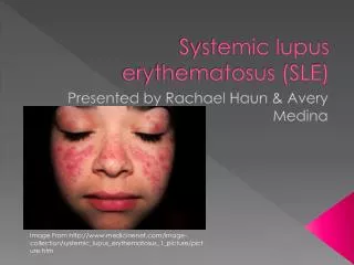
SYSTEMIC LUPUS ERYTHEMATOSUS + RIGHT KNEE JOINT PAIN. Ramos, Ronald Rangel, Erika Raymundo , Nikko Rayos , Karen Recio , Maria Kristina Reyes, Carmen Reyes, Jenilene Reyes, Lourdes. Rivera, Laila Rivere , Djeaune Robosa,Dean Rodas , Francis Rodriguez, Shereen

E N D
SYSTEMIC LUPUS ERYTHEMATOSUS+ RIGHT KNEE JOINT PAIN Ramos, Ronald Rangel, Erika Raymundo, Nikko Rayos, Karen Recio, Maria Kristina Reyes, Carmen Reyes, Jenilene Reyes, Lourdes Rivera, Laila Rivere, Djeaune Robosa,Dean Rodas, Francis Rodriguez, Shereen Rogelio, Ma.Graciela Roque, Marianne D2- MEDICINE 2011
General Data 16 y/o Female Catholic Student
Outline of Presentation To present a case of 16 y/o F with SLE nephritis with acute monoarticular arthritis, RKJ To present differential diagnosis of monoarticular arthritis To present diagnostic work-up of monoarticular arthritis To present treatment algorithm of monoarticular arthritis Prognosis as monoarticular arthritis
Chief Complaint Right Knee Joint Pain
16 F: SLE nephritis + RKJ Pain History of Present Illness
16 F: SLE nephritis + RKJ Pain History of Present Illness
16 F: SLE nephritis + RKJ Pain History of Present Illness
16 F: SLE nephritis + RKJ Pain History of Present Illness ADMISSION
16 F: SLE nephritis + RKJ Pain Review of Systems No pruritus, Jaundice or rashes No eye discharge, redness or pain, (+) photosensitity No nasal discharge, epistaxis No aural discharge, tinnitus or deafness (+) aphthous ulcer on left labial commissure, (+) periangular stomatitis No sore throat, no neck stiffness No breast mass or discharge No cough or colds No cyanosis, palpitations, fainting spells or orthopnea No dysuria, frequency or incontinence No abdominal pain, vomiting, diarrhea or constipation No polyuria, polyphagia, polydipsia or heat/cold intolerance No abnormal bleeding or bruising No loss of consciousness or sleeping difficulty
16 F: SLE nephritis + RKJ Pain Past Personal History • Feeding History • Typically: Snacks: fruits (banana) Meals: 1 cup rice + fish and vegetables Beverage: • June 2009 diarrheal episode: • Street food from school: buko, corn in a cup • Dinner: egg, cabbage • HEEADSSS H close relationship with family E 2nd year HS, good grades, frequent absences E well-balanced diet A school work, watch movies D denies use of illicit drugs S good relationship with the opposite sex; no sexual contact S no suicidal ideation S no extreme activities
16 F: SLE nephritis + RKJ Pain Past Personal History • Menstrual History M June 2009 I - D 3 days A 3 pads/day S dysmenorrhea • Immunization History (+) BCG* - 2 doses (+) Hep B* - 3 doses (+) OPV* - 3 doses (+) DPT* - 3 doses (+) Measles – 1 dose (-) MMR (-) Hib (-) booster dose for DTP and Polio *all were given at a local health center. Unrecalled dates. No document was presented.
16 F: SLE nephritis + RKJ Pain Family History (-) Asthma, DM, heart diseases, TB, seizure, blood dyscrasia, stroke, arthritis (+) HPN – mother’s side (+) SLE – patient’s twin sister Socioeconomic and Environmental History Father – breadwinner (maintenance personnel) Mother – housewife; primary health care giver Family of 6, living in a rented 1-bedroom bungalow made of concrete; not well-ventilated; area prone to flooding Drinking water source – from the tap, boiled Garbage – segregated; regular collection Pet – cat; contact with strays Second hand smoke – no exposure
16 F: SLE nephritis + RKJ Pain Past Medical History 2003 (PCMC) SLE: [polyarthritis, photosensitivity, anemia, rash, oral ulcers, nephritis + serology] Rx : prednisone, ASA, CaCO3 + Vit D Lupus Nephritis, Class IV -> Cyclophosphamide treatment x 11 cycles 2007 (USTH) LJP 394 clinical trial -> Abetimus 900 mg Rx : prednisone, ASA, CaCO3 +Vit D, enalapril May 2009 1st CyPT Rx : prednisone, CaCO3 +Vit D, enalapril
OPD LAB TESTS (June 24) Fecalysis brown, watery RBC = 0/hpf WBC = 0/hpf bacteria = 4+ no ova seen Urinalysis yellow, slightly turbid sp grav = 1.015 pH = 5.0 albumin = 4+ sugar = neg RBC = >100/hpf WBC = 40-70/hpf mucus threads = 2+ 16 F: SLE nephritis + RKJ Pain • CBC Hb = 14.1 g/dL Hct = 45 % WBC = 11.2 x 10⁹ cells/L bands = 1 segmenters = 90 lymphocytes = 6 monocytes = 3 Platelets = adequate • Serum Creatinine = 1.19 mg/dL
16 F: SLE nephritis + RKJ Pain PHYSICAL EXAMINATION ON ADMISSION (June 29) Conscious, coherent, wheelchair borne, not in cardiorespiratory distress BP 120/60 PR 100 bpm,regular RR 22 cpm,regular Wt. 30.9 kg (<p5) Ht. 139 cm (<p5) T 37C Normocephalic, normal hair distribution, (+) moon facies Pink palpebral conjunctiva, anicteric sclera, EOM full and equal, 2-3 mm ERTL (-) tragal tenderness, nonhyperemic EAC, intact tympanic membrane, AU, (-) aural discharge
16 F: SLE nephritis + RKJ Pain PHYSICAL EXAMINATION ON ADMISSION (June 29) Midline nasal septum, (-) nasal discharge, turbinates not congested Moist buccal mucosa, uvula midline, nonhyperemic PPW, (-) dental caries, (+) oral ulcers, (+) periangular stomatitis L (+) Buffalo hump, supple neck, (-) palpable cervical lymph nodes No chest wall deformities, trachea midline, symmetric chest expansion, resonant on percussion, normal vocal and tactile fremiti, clear breath sounds
PHYSICAL EXAMINATION ON ADMISSION (June 29) Adynamic precodium, AB at 5th LICS MCL, (-) murmurs, heaves, thrills or lifts Flat abdomen, midline umbilicus, normoactive bowel sounds, soft, nontender, no organomegaly Extremities: (-) bipedal edema RKJ=(+) pain, effused, warm, tender; limited passive and active range of motion good peripheral pulses 16 F: SLE nephritis + RKJ Pain
16 F: SLE nephritis + RKJ Pain Salient Features 16 y.o./Female diagnosed with SLE nephritis class IV (2003) (+) aphthous ulcer on left labial commissure, (+) periangular stomatitis, (+) Photosensitivity RKJ=(+) pain, effused, warm, tender; limited passive and active range of motion Watery, non-mucoid, non-bloody diarrhea 4x/day, 200cc /episode History of eating street foods BP 120/60 (+) HPN – mother’s side (+) SLE – patient’s twin sister
16 F: SLE nephritis + RKJ Pain Working Diagnosis SLE in flare (renal, hematologic, mucocutaneous) Monoarticular Arthritis (RK), etiology to be determined
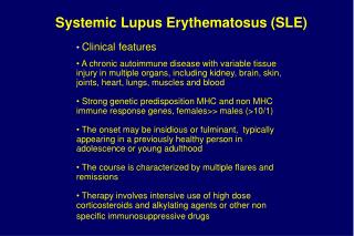
16 F: SLE nephritis + RKJ Pain Diagnostic Criteria for SLE ≥4 of 11 criteria is needed to diagnose SLE. Specificity is ~95% and sensitivity is ~75% • Malar or discoid rash • Photosensitivity • Oral ulcers • Arthritis • Serositis • Renal Disorder • Neurologic disorder • Hematologic disorder • Immunologic disorder • Antinuclear antibodies Fauci, et al. Harrison’s Principles of Internal Medicine 17th ed.
16 F: SLE nephritis + RKJ Pain Predisposing Factors for the Development of SLE http://www.hss.edu/conditions_14138.asp Fauci, et al., Harrison’s Principles of Internal Medicine. 17th ed. Vol. II. Female gender Genetics Susceptibility genes such as homozygous deficiencies of early components of complement (C1q,r,s; C2; C4) Environment Autoantibodies
16 F: SLE nephritis + RKJ Pain Lupus Nephritis Fauci, et al., Harrison’s Principles of Internal Medicine. 17th ed. Vol. II. Page 2077 Common and serious complication of SLE Clinical signs of renal disease present in the patient are proteinuria, hematuria and hypertension and decreased renal function. Patient has Class IV: Diffuse Lupus Nephritis
Classification of Lupus Nephritis (International Society of Nephrology and Renal Pathology Society) 16 F: SLE nephritis + RKJ Pain Class I: Minimal Mesangial Lupus Nephritis Class II:Mesangial Proliferative Lupus Nephritis Class III: Focal Lupus Nephritis Class IV: Diffuse Lupus Nephritis • Class IV-S (A) • Class IV-G (A) • Class IV-S (A/C) • Class IV-G (A/C) • Class IV-S (C) • Class IV-G (C) Class V: Membranous Lupus Nephritis Class VI: Advanced Sclerotic Lupus Nephritis
Differential Diagnosis 16 F: SLE nephritis + RKJ Pain R. Knee Pain Hx, PE ARTICULAR NON-ARTICULAR INFLAMMATORY NON-INFLAMMATORY INFECTIOUS CRYSTAL-INDUCED IMMUNE-RELATED REACTIVE IDIOPATHIC SA, Culture BACTERIAL VIRAL FUNGAL Fauci, et al. Harrison’s Principles of Internal Medicine 17th ed.
16 F: SLE nephritis + RKJ Pain Laboratory Exams http://emedicine.medscape.com/article/1268369-overview Harrison’s Principle of Internal Medicine, 16th edition 2005 • CBC, WBC and differential count • To evaluate whether patient’s overall health status • to find anemia and an infection • WBC count higher than 12,000/µL seen in septic arthritis • Blood GS/CS • To determine whether bacteremia is present and the microorganism in
16 F: SLE nephritis + RKJ Pain Laboratory Exams • Urinalysis • Creatinine • Electrolytes: Na K
16 F: SLE nephritis + RKJ Pain Chest X Ray
16 F: SLE nephritis + RKJ Pain Laboratory Exams Harrison’s Principle of Internal Medicine, 17th edition 2008 • Synovial fluid cell count, GS/CS and AFB • most important laboratory test to detect septic arthritis • Synovial cell counts seen in bacterial arthritis: • average of 100,000/L (range, 25,000–250,000/L) • with >90% neutrophils
16 F: SLE nephritis + RKJ Pain Arthrocentesis • Joint aspiration using a sterile needle and syringe to drain fluid from the joint • To establish the cause of an acute monoarthritis or polyarthritis • A therapeutic procedure • to drain large effusions or hemarthroses • to instill corticosteroids local anesthetic - “Arthrocentesis of the Knee”, n engl j med 354;19
Laboratory Tests that were not done 16 F: SLE nephritis + RKJ Pain • Blood gas • since RR of patient = 32 cpm • BUN Creatinine Ratio • to determine if the condition is pre-renal (increased protein catabolism, corticosteroid therapy), renal (acute or chronic renal failure) or post-renal (Urinary tract obstruction) • normally 10:1 to 20:1 emedicine.medscape.com/article/1268369-overview www.cfp.ca/cgi/content/full/55/4/374
Laboratory Tests that were not done 16 F: SLE nephritis + RKJ Pain Harrison’s Principle of Internal Medicine, 16th edition 2005 • ESR and CRP • markers for inflammation and it will be useful in detecting if patient is responding to the therapy • not specific • elevated with infection, inflammation • Normal values: • ESR (females): 0-25 mm/hr • CRP 0.08-3.1 mg/L
Imaging studies 16 F: SLE nephritis + RKJ Pain Plain X-ray of Joint • To see if there’s synovial thickening and effusion (it distends the joint – to see if it needs drainage) • To see how much joint space is involved (joint narrowing) • Demineralization (induces hyperemia and immobilization) • If the cartilage is already destroyed or if there’s already joint destruction(30-70 cases per 100,000 in patients with autoimmune disease)
Therapeutic Plan Relieve pain and lessen effusion in the joint Identify the microorganism and treat sepsis Prevent SLE flare Maintain full mobility of joint
Therapeutic Plan: SLE Nephritis 16 F: SLE nephritis + RKJ Pain Fauci, A. et al. (2008) Harrison’s Principles of Internal Medicine. USA: McGraw –Hill Publishing Inc. • Systemic glucocorticoids • Prednisone • Cytotoxic or immunosuppressive agents • Cyclophosphamide • BSA x 0.75-1g of cyclophosphamide • ACE inhibitors • Enalapril • Calcium plus Vitamin D
Day 1 June 29 (Admission) Medications / Intervention Laboratory Exams 16 F: SLE nephritis + RKJ Pain Course in the ward Blood C/S CXR: Normal Arthrocentesis and lavage RKJ cefazolin 1g q8h IV • CBC • RBC 141, WBC 11.2 (N-94%, L 6%), plt adequate • Urinalysis • alb4+, sug neg, RBC>100, WBC- 40-70 • Fecalysis- bacteria 4+ • Crea- 1.19mg/dl • Synovial Fluid analysis • SF C/S • SF: 24cc, purulent • AFB and Gram’s stain: (-) • WBC=65,600/cumm (Neutro: 89, lymphos 11) amikacin500mg IV q10h SF Culture: Gram neg bacilli after 13H incubation
Day 2 June 30 Medications / Intervention Laboratory Exams 16 F: SLE nephritis + RKJ Pain Course in the ward Crea-0.5 mg/dL, Na-170 K-3.2 • Urinalysis: • lt yellow, sl turbid • ph 6, SG 1.015, alb 2(+), sugar (-) • cast (Hyal 5/cs), • RBC 18-22/hpf, • WBC 8-10/hpf Arthrocentesis RKJ: 5cc amikacin500mg IV q10h prednisone60mg/d enalapril5mg/tab OD CaCO3+ Vit D 500mg/tab 1 tab OD
Day 3 July 1 Medications / Intervention Laboratory Exams 16 F: SLE nephritis + RKJ Pain Course in the ward SF C/S : Salmonella typhii sensitive to: ampicillin, TMX-sulfamethoxazole, ceftriaxone, cefotaxime, chloramphenicol chloramphenicol 500 mg/cap oral amikacin500mg q10 IV ampicillin-sulbactam1.5g q6 prednisone60mg/d enalapril5mg/tab OD CaCO3+ Vit D 500mg/tab 1 tab OD Arthrocentesis RKJ: 24 cc
Day 4 July 2 Day 5 July 3 Medications / Intervention Laboratory Exams 16 F: SLE nephritis + RKJ Pain Course in the ward Blood C/S: Salmonella typhii Blood C/S (+) Gm neg bacilli chloramphenicol 500 mg/cap oral Arthrocentesis RKJ: 15 cc prednisone60mg/d enalapril5mg/tab OD CaCO+ Vit D 500mg/tab 1 tab OD
Day 6 July 4 Day 7 July 5 Medications / Intervention Laboratory Exams 16 F: SLE nephritis + RKJ Pain Course in the ward none none chloramphenicol 500 mg/cap prednisone60mg/d enalapril5mg/tab OD CaCO3+ Vit D 500mg/tab 1 tab OD
Day 8 July 6 Day 9 July 7 Day 10 July 8 Day 11 July 9 Medications / Intervention Laboratory Exams 16 F: SLE nephritis + RKJ Pain Course in the ward • CBC: • Hb 106 • Plt 315 • WBC 12.6 • neutro 93, lympho5, • mono 2 Mini-arthrotomy of RKJ Improved and stable prednisone,enalapril, CaCO3+VitD, chloramphenicol Discharged
Fever Temperature Graph 16 F: SLE nephritis + RKJ Pain
Treatment • Arthrocentesis • Empiric IV therapy • Definitive therapy: • after identification of pathogen and antibiotic susceptibility tests • chloramphenicol for Salmonella typhii • Mini-Arthrotomy • Glucocorticoids and Calcium carbonate with vitamin D for SLE maintenance
16 F: SLE nephritis + RKJ Pain Arthrotomy Means an incision into a joint Used to remove loculations and debride infected synovium, cartilage, or bone
16 F: SLE nephritis + RKJ Pain Septic Arthritis http://www.hmc.psu.edu/healthinfo/s/septicarthritis.htm most commonly affects weight-bearing joints such as the knee Risk factor is the diagnosis of SLE in the patient The bacteria can reach the joint through hematogenous route from infection in other areas of the body
Septic Arthritis 16 F: SLE nephritis + RKJ Pain Synovial proliferation Enter joint space Formation of Panus thrombosis
Salmonella typhi family Enterobacteriaceae gram-negative enteric bacillus motile, facultative anaerobe susceptible to various antibiotics grows on MacConkey and EMB agars non-lactose fermenting no gas produced on TSI media differentiates it from other Enterobacteriaceae 16 F: SLE nephritis + RKJ Pain Harrison’s Principle of Internal Medicine, 17th edition 2008