Download
1 / 21
330 likes | 2.7k Vues
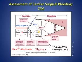
Hemostasis, Surgical Bleeding, and Transfusion. Patrick Chidi Obasi. Hemostasis. Complex process that prevents blood loss from disrupted intravascular space. Major physiologic events: Primary hemostasis Vascular constriction Platelet plug formation Secondary hemostasis Fibrin Formation

E N D
Hemostasis, Surgical Bleeding, and Transfusion Patrick Chidi Obasi
Hemostasis • Complex process that prevents blood loss from disrupted intravascular space. • Major physiologic events: • Primary hemostasis • Vascular constriction • Platelet plug formation • Secondary hemostasis • Fibrin Formation • Fibrinolysis
Vascular constriction • Initial vascular response to injury • Linked to platelet plug formation • Thromboxane A2 (TxA2), Serotonin, 5-hydroxytryptamine (5-HT), Bradykinin
Platelet Plug Formation • Conversion of cyclooxygenase to TXA2=> stimulates platelet motility and binding (aggregation) • Activated platelets adhere to fibrinogen via GP IIa/IIIb. • Plts adhere to von Willebrand factor on exposed collagen via glycoprotein (GP) Ia/IIa receptor • Adhesion results in the release of agonist like epi, collagen, thrombin, ADP, Serotonin • Platelet cyclooxygenase irreversibly inhibited by ASA and reversibly by NSAIDs
Coagulation/Fibrin Formation • Extrinsic pathway • Tissue factor (injured cells) + factor VII • Activates factors X and V • Converts prothrombin to thrombin • Thrombin then converts fibrinogen to fibrin • Elevated PT associated with abnormal extrinsic pathway cascade
Coagulation/Fibrin Formation • Intrinsic pathway • All components intrinsic to the circulating plasma • HMW kininogen + factor XII (Hageman factor) bind to exposed collagen • Activation of factors XI, IX + VIII => X+V • Convert prothrombin (factor II) to thrombin • Thrombin cleaves fibrinogen to fibrin • Abnormal measured by a PTT
Fibrinolysis • Plasminogen level known to rise as a consequence of exercise, occlusion, and anoxia • tPA-released from endothelium converts plasminogen to plasmin • Plasmin degrades factors V and VIII, fibrinogen and fibrin-leading to lose plt plug
Notes on Coagulation Factors • Factor II aka prothrombin • Factor III aka antithrombin • Heparin binds to AT III • Deficiency results in spontaneous thrombosis • Treat def with FFP (has AT-III) and then heparin
Notes on Coagulation Factors • Factors V-labile factor • Factor VI – shortest half life. Begins extrinsic pathway • Factor VIII – labile factor -- only factor not synthesized in liver (made in endothelium) • Factor X – Convergence point for both pathways • Factor XII – aka Hageman factor. Begins intrinsic pathway • Factor XIII – Helps crosslink fibrin
Local Hemostasis • Goal is to prevent or interrupt the flow of blood from a disrupted vessel that has been incised or transected • Digital pressure effective and has adv. of being less traumatic • Hemostat, hemoclips, transfixion sutures (prevents slipping) • The adventitia and media constitute the major holding forces in a vessel wall
Local Hemostasis • Thermal agents: Harmonic scalpel, electrocautery bovie (causes collagen denaturing leading to coagulation) • Direct cooling with iced saline causes vessels to undergo necrosis by dehydration and destruction of lipid molecules • Chemical agents: epi (induces vasoconstriction), gel foam, surgicel, topical thrombin, fibrin sealant)
Heparin Therapy • Potentiates action of AT III • Inhibited by admin of protamine (1mg of protamine:100 units of heparin for reversal) • Measured by aPTT • Want PTT between 60-90 for anticoagulation • Does not cross placental barrier • Stop 4-6 hours before surgery
HIT/HITTS • Due to antiplatelet antibodies (IgG) that results in plt destruction • Can cause plt aggregation and thrombosis (“white clot”) • Typically seen 5 to 7 days in first exposure • May occur within 1 to 2 days in re-exposure • Suspected if plt count falls more than 100k or drops by 50% from baseline • LMWH (Lovenox) still at low risk • Tx: stop heparin, start lepirudin/Hirudin, argatroban, dextran
Coumadin Therapy • Inhibits cyclo-oxygenation (decarboxylation) step in Vit K synthesis of coag • Effects reversed by Vit K admin • Stop 7 days before surgery • Measured by INR • INR >1.5 – contraind. for surgery • INR >1.3 – contraind. for central line placement, perc. needle bx and eye surgery • Teratogenic
Warfarin-induced skin necrosis • Occurs when pt is placed on coumadin without heparin first • Due to short ½ of prot. C and S, which are first to decrease in levels compared with the procoagulants (factors II, VII, IX and X) • Above results in relative hyperthrombotic state • Tx: Heparin if it occurs, prevent by heparinizing first
Hypercoagulability • Leiden Factor • Resistance to activated prot C and S • Spont venous thrombosis • Most commoncongenital hypercoagulability disorder • Tx: heparin, warfarin • Polycythemia vera • Defect in plt function • Can have thrombosis/bleeding • Keep Hct<48 and plts <400 before surgery • Tx: ASA
Hypercoagulability • AT-III • Pts have spont venous thrombosis • Heparin does not work • Tx: AT-III conc, FFP then followed by Heparin
Hypercoagulability • Lupus Anticoagulant (Procoagulant) • Antiphospholipid antibodies • Prolonged PTT in the face of hypercoag state • Dx: Prolonged PTT not corrected by FFP • Tx: Heparin, coumadin • Acquired hypercoag: Tobacco, malignancy, IBD, infections, post-op state
DVT • Stasis, venous injury, hypercoagulability • Risk factors include cancer (#1), obesity, varicose veins, history of DVT, immobility • Most common site in post-op pt is the pop vein • Activated prot C resistance due to factor V leiden def is the most common cause of idiopathic recurrence • Dx/Tx:
PE • Presents with SOB, tachycardia, hypoxemia, normal or decreased PaCo2 • Most common from above knee (iliofemoral region) • Dx/Tx • Indication for surgical procedure • When filters are indicated • Pt who have undergone pulm emboletomy • Pts with contraindication for anticoagulation • PE while on anticoagulant • Free floating above knee DVT
PRBC Storage • 1 Unit of PRBC has a vol of 250 ml • Storage life is about 35 days • ↑K+, ↓ pH, ↓ 2.3-DPG level (↓ O2 del capacity), ↑ IL-1, and IL-6 • Each unit expected to raise H/H by 1gm/3% • Fever without hemolysis is the most common transfusion reaction • Acute hemolysis is the most common cause of transfusion related death • Hypotension, shock, dyspnea, bronchospasm, dizziness and flushing are sign of hemolysis • Tx: Supportive