Download
1 / 9

E N D
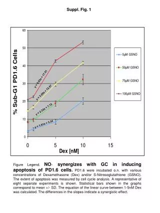
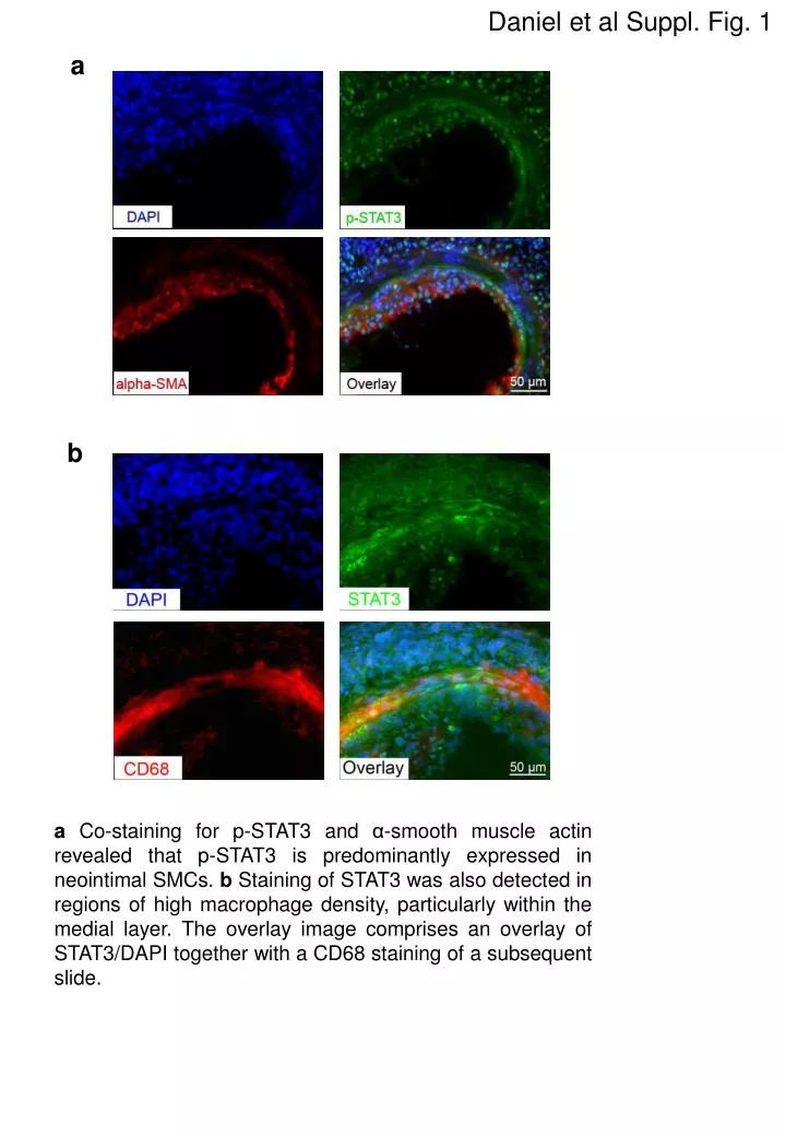
Daniel et al Suppl. Fig. 1 a b aCo-staining for p-STAT3 and α-smooth muscle actin revealed that p-STAT3 is predominantly expressed in neointimal SMCs. b Staining of STAT3 was also detected in regions of high macrophage density, particularly within the medial layer. The overlay image comprises an overlay of STAT3/DAPI together with a CD68 staining of a subsequent slide.
Daniel et al Suppl. Fig. 2 a b * * STAT3 STAT3 mRNAexpression Tubulin FCS - 8h 12h FCS - 4h 8h a+b In stimulated SMCs, STAT3 expression was foundtobeup-regulatedon themRNAlevelat 4 and 8 hoursand on theproteinlevelat 8 and 12 hours after stimulationusing real-time PCR and Western blotting , respectively (*P<0.05, n=4).
Daniel et al Suppl. Fig. 3 b a survivin cyclin D1 * * a+b Real-time PCR showed a significant up-regulation of cyclin D1 and survivn mRNA levels in the dilated artery at 3 weeks after dilation (*P<0.05, n=4).
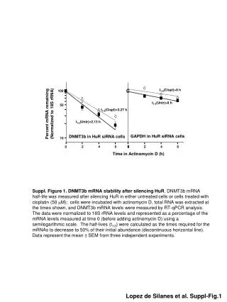
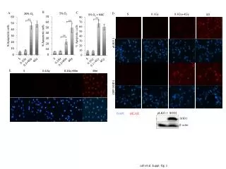
Daniel et al Suppl. Fig. 4 a * * FCS WP1066 (µM) b * * * FCS WP1066 (µM) aSMCs were incubated in basal medium or growth medium supplemented with FCS in the absence or presence of different concentrations of WP1066 for 24 h. Apoptosis of SMCs was evaluated by a TUNEL-based cell death detection ELISA (*P<0.05, n=4).b SMCs wereincubatedwithgrowth medium in theabsenceorpresenceof different concentrationsof WP1066, andthefraction non-necroticcells was determinedbytrypanblueexclusion (*P<0.05, n=4).
Daniel et al Suppl. Fig. 5 a b Index ofre-endothelialization (n=0-6) aRepresentativecrosssectionsoffemoralarteriesfromcontrolmice (left) ormicetreatedwith WP1066 (right) stainedfor CD31 (PECAM-1) at 3 weeks after dilation. b Re-endothelialization was determined by estimating the lumen coverage on a scale of 0-6 (0, no coverage; 6, complete coverage) (n=6, P=n.s.).
Daniel et al Suppl. Fig. 6 a uninjuredcontrol uninjured WP1066 c b 50µm 50µm Uninjured WP1066 vWF uninjured WP1066 TUNEL aRepresentative en facestainingwith Evans blueofuninjuredfemoralarteriesfromcontrols (left) or after localapplicationof WP1066 (right). b The integrityoftheendotheliallayer after applicationof WP1066 was also confirmed by staining for endothelial markers (arrowhead indicates staining for von-Willebrand-factor).c Apoptotic cell death was very rare after application of WP1066 to uninjured arteries and occurred mainly in the adventitia and neighboring tissue but not within the intima or media (arrow indicates TUNEL-positive cell in the neighboring tissue).
Daniel et al Suppl. Fig. 7 a b d c f e g h
i j l k n m o p
q a-q Blood testswereperformedat 1 week after injury, in order toanalyzethesystemiceffectsof WP1066. There was nosignificantdifferencebetweenvehicle (DMSO) and WP1066 treatedmice.