Download
1 / 19
190 likes | 200 Vues
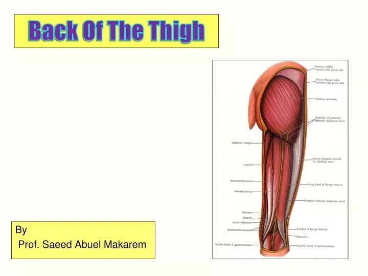
Back Of The Thigh. By Prof. Saeed Abuel Makarem. Cutaneous innervations of the back of thigh: 1- Posterior Cutaneous nerve of thigh,S1,2 & 3. 2- Posterior branch of lateral cutaneous nerve of thigh. 3- Posterior branch of medial cutaneous nerve of thigh.

E N D
Back Of The Thigh By Prof. Saeed Abuel Makarem
Cutaneous innervations of the back of thigh: • 1- Posterior Cutaneous nerve of thigh,S1,2 & 3. • 2- Posterior branch of lateral cutaneous nerve of thigh. • 3- Posterior branch of medial cutaneous nerve of thigh.
The back of thigh contains the following: Hamstrings muscles. Sciatic nerve. Chain of anastomosis (perforating arteries & cruciate anastomosis.
You should remember the following: • The hamstrings are the main Flexors of the knee, but they are also, Extensors of the hip. • All hamstrings arise from Ischial tuberosity, ExceptShort head of biceps from linea aspera and lateral supracondylar ridge of the femur. • Biceps femoris inserted into head of the Fibula. • Semimembranosus inserted into a groove in the back of medial tibial condyle. Some fibers from the semimembranosus pass upward and laterally forming oblique popliteal ligament of the knee.
Semitendinosus & long head of biceps have a common origin from the Ischial tuberosity. • Semitendinosus inserted into upper part of medial surface of tibia (SGS( • Muscles inserted into tibia rotate leg medially. • Muscle inserted into fibula rotates leg laterally. • All hamstrings are supplied by tibial nerve (medial popliteal) except short head of biceps by common peroneal nerve (lateral popliteal).
Sciatic nerve • Larger of the two terminal branches of sacral plexus. • Root value: • L4 and 5 ; S1, 2 and 3. • Leaves the pelvis through greater sciatic foramen, below piriformis. • Covered by gluteus maximus. • It crosses the following from above downwards • Tendon of obturator internus and 2 gemelli, Quadratus femoris, adductor magnus • Surface anatomy: draw a line which begins from midpoint between greater trochanter & ischial tuberosity to the apex of popliteal fossa.
Popliteal Fossa • Definition: Diamond shaped space in the back of the knee joint. • Boundaries: • Upper lateral: Biceps femoris. • Upper medial: Semitendinosus, semimembranosus, supplimented by sartorius, gracilius and Ischial part of adductor magnus. • Lower medial: medial head of gastrocnemius. • Lower lateral: Lateral head of gastrocnemius and Plantaris. • Lower limit of the fossa is the distal border of popliteus. Roof: Skin, superficial fascia containing upper part of small saphenous vein, deep fascia (popliteal fascia).
Surface anatomy of Right popliteal fossa Upper medial border (1) (Semitendinosus(2), semi- membranosus, Ischial part of adductor magnus, gracilius & sartorius. Lower medial (3): medial head of gastrocnemius Lower lateral (5) Lateral head of gastrocnemius & Plantaris. Upper lateral : Biceps femoris(6). 4- Soleus muscle.
Floor of popliteal fossa: From above downwards: Popliteal surface of the femur Back of capsule of knee joint, Fascia covering popliteus muscle. Contents: Medial popliteal nerve (Tibial). Lateral popliteal nerve (Common peroneal) Popliteal artery. Popliteal vein. Popliteal lymph nodes. Popliteal fat.
Popliteal Artery • Origin: • It is the continuation of femoral artery at the opening of adductor magnus. • Termination: • At the distal border of popliteus it divides into anterior & posterior tibial arteries. • Branches: • 1- muscular branches. • 2- Genicular branches (Superior lateral, superior medial, inferior lateral, inferior medial & middle genicular arteries) • It is the deepest structure in the fossa as it runs on the floor of the fossa.
Medial Popliteal Nerve • Is the larger of two terminal branches of sciatic nerve, which bisects the fossa. • It is the most superficial structure in the fossa • Crossing popliteal vessels superficial from lateral to medial. • Branches: • 1- Cutaneous: Sural nerve. • 2- Muscular: (5) To each head of gastrocnemius, popliteus, Plantaris and soleus. • 3- Articular: (3) Superior medial, inferior medial, and middle genicular nerves. • Termination: At the distal border of popliteus it is continuous as posterior tibial nerve.
Lateral popliteal(common Peroneal) Nerve • Smaller of the 2 terminal branches of sciatic. • Enters the fossa through its upper angle. • Runs close to medial side of the tendon of biceps. • Leaves the fossa through its lateral angle. • It crosses Plantaris & lateral head of gastrocnemius. • It curves on the lateral side of fibular neck. • Termination: Within the substance of peroneus longus it divides into 2 terminal branches: • anterior tibial (deep peroneal) and • musculo-cutaneous , ( superficial peroneal).
Branches: • 1- Cutaneous: 2 Lateral Cutaneous nerve of the calf. Sural communicating nerve • 2- Muscular: No muscular branches in the fossa. • 3- Articular: (3) Superior lateral genicular nerve. Inferior lateral genicular nerve. Recurrent genicular nerve.
Popliteal Vein • Begins at the distal border of popliteus muscle by the union of the 4 venae comitantes of the anterior & posterior tibial arteries. • Leaves the fossa at its upper end as it becomes the femoral vein at the opening of adductor magnus. • Tributaries: muscular, 5 genicular and small sphenous vein. • It lies deep to popliteal nerves and superficial to the popliteal artery.