Download
1 / 48
530 likes | 1k Vues
Cell Division and Mitosis. Chapter 10. 10.1 The Cycle of Cell Growth and Division: An Overview. The products of mitosis are genetic duplicates of the dividing cell Chromosomes are the genetic units divided by mitosis. Mitotic Cell Division. DNA replication

E N D
Cell Division and Mitosis Chapter 10
10.1 The Cycle of Cell Growth and Division: An Overview • The products of mitosis are genetic duplicates of the dividing cell • Chromosomes are the genetic units divided by mitosis
Mitotic Cell Division • DNA replication • Equal separation (segregation) of replicated DNA molecules • Delivery to daughter cells • Two new cells, same information as parent cell
Mitosis • Mitosis is the basis for • Growth and maintenance of body mass in multicelled eukaryotes • Reproduction of many single-celled eukaryotes
Chromosomes • DNA of eukaryotic cells is divided among individual, linear chromosomes • Located in cell nucleus • Ploidy of a cell or species • Diploid (2n) • Haploid (n)
Eukaryotic Chromosomes Fig. 10-2, p. 203
Sister Chromatids • DNA replication and duplication of chromosomal proteins produces two exact copies (sister chromatids) • Chromosome segregation occurs during cell division
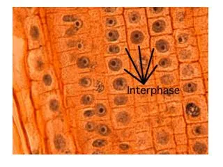
10.2 The Mitotic Cell Cycle • Interphase extends from the end of one mitosis to the beginning of the next mitosis • After interphase, mitosis proceeds in five stages • Cytokinesis completes cell division by dividing the cytoplasm between daughter cells
10.2 (cont.) • The mitotic cell cycle is significant for both development and reproduction • Mitosis varies in detail, but always produces duplicate nuclei
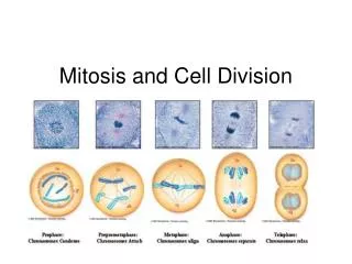
Mitotic Cell Cycle • Includes mitosis and interphase • Mitosis occurs in five stages • Prophase • Prometaphase • Metaphase • Anaphase • Telophase
The Cell Cycle Fig. 10-3, p. 203
Interphase Fig. 10-4a (1), p. 204
Stage 1: Prophase • Chromosomes condense into short rods • Spindle forms in the cytoplasm
Prophase Fig. 10-4a (2), p. 204
Stage 2: Prometaphase • Nuclear envelope breaks down • Spindle enters former nuclear area • Sister chromatids of each chromosome connect to opposite spindle poles • Kinetochore of each chromatid attaches to the spindle microtubules
Prometaphase Fig. 10-4a, p. 204
Spindle Connections at Prometaphase Fig. 10-6, p. 206
Stage 3: Metaphase • Spindle is fully formed • Chromosomes align at metaphase plate • Moved by spindle microtubules
Metaphase Fig. 10-4b, p. 204
Stage 4: Anaphase • Spindle separates sister chromatids and moves them to opposite spindle poles • Chromosome segregation is complete
Anaphase Fig. 10-4b, p. 204
Stage 5: Telophase • Chromosomes decondense • Return to extended state typical of interphase • New nuclear envelope forms around chromosomes
Telophase Fig. 10-4b, p. 204
Mitosis Fig. 10-5, p. 206
Cytokinesis • Division of cytoplasm completes cell division • Produces two daughter cells • Each daughter nucleus produced by mitosis
Cytokinesis in Animal Cells • Proceeds by furrowing • Band of microfilaments just under the plasma membrane contracts • Gradually separates cytoplasm into two parts
Cytokinesis by Furrowing Fig. 10-8, p. 208
Plant Cytokinesis • Cell wall material is deposited along the plane of the former spindle midpoint • Deposition continues until a continuous new wall (cell plate) separates daughter cells
Cytokinesis by Cell Plate Formation Fig. 10-9, p. 208
10.3 Formation and Action of the Mitotic Spindle • Animals and plants form spindles in different ways • Mitotic spindles move chromosomes by a combination of two mechanisms
Spindle Formation • In animal cells • Centrosome divides, the two parts move apart • Microtubules of the spindle form between them • In plant cells with no centrosome • Spindle microtubules assemble around the nucleus
Centrosome and Spindle Formation Fig. 10-10, p. 210
In the Spindle • Kinetochore microtubules • Run from poles to kinetochores of chromosomes • Nonkinetochore microtubules • Run from poles to a zone of overlap at the spindle midpoint without connecting to chromosomes
A Fully Developed Spindle Fig. 10-11, p. 210
During Anaphase • Kinetochores move along kinetochore microtubules • Pulling chromosomes to the poles • Nonkinetochore microtubules slide over each other • Pushing the poles farther apart
Anaphase Spindle Movements Fig. 10-12, p. 211
Kinetochore Movement Fig. 10-13, p. 211
10.4 Cell Cycle Regulation • Cyclins and cyclin-dependent kinases • Internal controls that directly regulate cell division • Internal checkpoints • Stop cell cycle if stages are incomplete • External controls • Coordinate mitotic cell cycle of individual cells within overall activities of the organism
Cell Cycle Control (1) • Complexes of cyclin and a cyclin-dependent protein kinase (CDK) • Directly control cell cycle • CDK • Is activated when combined with a cyclin • Adds phosphate groups to target proteins, activating them
Cell Cycle Control (2) • Activated proteins trigger the cell to progress to the next cell cycle stage • Each major stage of the cell cycle • Begins with activation of one or more cyclin/CDK complexes • Ends with deactivation of complexes by breakdown of cyclins
Cyclin/CDK Control Fig. 10-15, p. 214
Internal Controls • Important internal controls create checkpoints • Ensure that the reactions of one stage are complete before cycle proceeds to next stage
External Controls • Based on surface receptors that recognize and bind signals • Peptide hormones and growth factors • Surface groups on other cells • Molecules of the extracellular matrix • Binding triggers internal reactions that speed, slow, or stop cell division
Cancer • Control of cell division is lost • Cells divide continuously and uncontrollably • Form rapidly growing mass of cells that interferes with body functions • Cancer cells break loose from their original tumor (metastasize) • Form additional tumors in other parts of the body
Tumor Cells Fig. 10-16, p. 215
Animation: Mitosis overview PLAY ANIMATION