Download
1 / 6
540 likes | 3.8k Vues
Onion vs. Cheek Cell. Comparing and contrasting onion and cheek cells. Inquiry Question. How do you think an onion cell would be different from a cheek cell? Describe shape, size, and organelle differences that you expect to observe. Gather Materials. Tweezers Onion

E N D
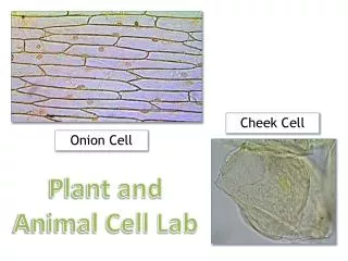
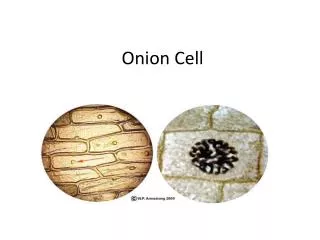
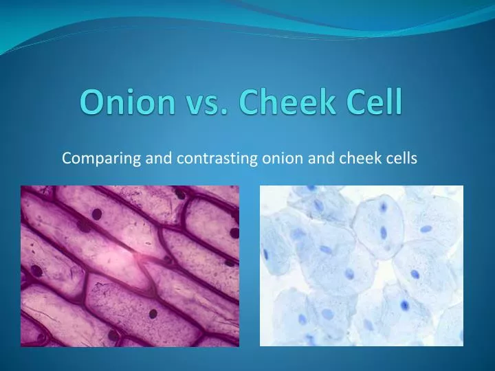
Onion vs. Cheek Cell Comparing and contrasting onion and cheek cells
Inquiry Question How do you think an onion cell would be different from a cheek cell? Describe shape, size, and organelle differences that you expect to observe.
Gather Materials • Tweezers • Onion • Slide and slip cover • Iodine • Dropper • Water • Microscope • Toothpick with blunt edge
How to prepare an onion and cheek cell slide • http://www.youtube.com/watch?v=GHnndVuaync&safety_mode=true&persist_safety_mode=1 Check out this youtube video for detailed instructions on how to prepare your cheek and onion cell for observation.
Procedure-onion cell 1. Peel the delicate transparent tissue from the inner surface of a piece of onion using tweezers. 2. Make a wet mount by placing the onion tissue, unwrinkled, in a small drop of water on a glass slide. 3. Add one small drop of iodine stain to the tissue and cover with a cover slip as directed. Put the edge of the cover slip at the edge of the microscope slide. Drag the cover slip to the edge of the water/iodine and drop it on top. 4. Examine the onion cells at low power, focus as necessary. 5. Next examine the cells at medium and high power. 6. Draw what you see inside the microscope. Label the parts including nucleus, cell membrane, cell wall, and nuclear membrane. 7. Answer the questions on your lab sheet about the onion cells.
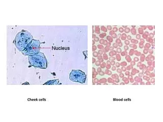
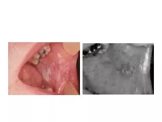
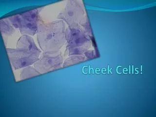
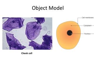
Procedure-cheek cell • 1. Place a drop of water on a clean slide. Gently scrape the inside of your cheek with the blunt end of a clean toothpick and stir the material on the toothpick in the drop of water on the slide. (Put toothpick in trash.) • 2. Add one small drop of iodine to the slide and then add a coverslip as directed above. • 3. Focus and examine the slide under low power before moving to the higher magnifications. • 4. Prepare a diagram showing 3 - 4 cells of the cheek and label structures you can identify. (Don't forget to identify magnification of the drawing.) • 5. Answer the cheek cell questions on your lab sheet.