Download

1 / 23
360 likes | 1.66k Vues
How to Diagnose and Assess Severity of Mitral Regurgitation by Echo. Noel Black Chief Cardiac Physiologist South Eastern Trust. Modalities. 2D and M-Mode Colour Doppler Pulsed Wave and Continuous Wave Doppler 3D. M-Mode. Left atrial dilatation Left ventricular dilatation

E N D
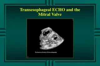
How to Diagnose and Assess Severity of Mitral Regurgitation by Echo Noel Black Chief Cardiac Physiologist South Eastern Trust
Modalities • 2D and M-Mode • Colour Doppler • Pulsed Wave and Continuous Wave Doppler • 3D
M-Mode • Left atrial dilatation • Left ventricular dilatation • Left ventricular volume overload pattern • Increased D-E amplitude of the mitral valve anterior leaflet
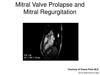
2-D • Status of the Mitral Valve Apparatus • Leaflet prolapse • Rheumatic disease • Myxomatous degeneration
Organic Aetiology • Calcification • Vegetation • Mass
Functional Ischaemic • Left ventricular impairment • Chordal/papillary muscle involvement
Functional. LV/LA dilatation • Mitral annular dilatation (normal 2.3+- 0.5cm) A4Ch view • LA dilatation • LV dilatation
Disease Process Aetiology Mechanism Non-Ischaemic Ischaemic Organic Rheumatic Ruptured PM Prolapse Endocarditis Flail leaflet Functional Cardiomyopathy Post-MI
Colour Flow Doppler • 1.Flow distribution (jet size) • 2.Vena contracta • 3.PISA
1.Flow distribution • How far the regurgitant jet extends into the LA. • Trace area of the jet and LA • Jet area (cm)2 • Severe MR: >10 • Jet area / LA (%) • Severe MR: >40
Colour Flow Doppler • Jet position in relation to the mitral leaflets. • Evidence of leaflet perforation. • Multiple or single jets
The direction of the regurgitant jet Centrally anteriorly posteriorly directed. Away from abnormal leaflet. Colour doppler
Consider Image quality • Poor image quality may underestimate severity
Consider –Jet direction Direction of the Jet (entrainment effect) • Central jet overestimated • Eccentric jet underestimated
Influence of Colour Gain Settings on Colour jet size -50-60 cm/s
2.Vena Contracta width • Narrowest region at the mitral valve level • 2 planes • Nyquist 50-60 cm/s • Zoom to optimise visualisation • Colour sector as narrow as possible • Maximal lateral and temporal resolution • Mild MR : • VC <0.3cm • Severe MR : VC >0.7cm
Consideration • VC width is inaccurate with multiple jets
Consideration • VC should not be measured in Apical 2Ch view • Parallel to the mitral orifice. • Overestimation.
3.Proximal Isovelocity surface area (PISA) • Hemishells • Flow convergence area • Increases with severity of regurgitation
Calculating (PISA) • Apical 4Ch view • Narrow sector width • Minimise depth • Zoom • Adjust Colour Doppler alaising velocity (20-40 cm/s)
PISA • Measure the radius of the hemisphere. (red/blue interface) • PISA radius =2πr2 (cm2) • Mild MR: <0.4 • Severe MR:>1.0
Consideration Non-circular orifice