Download

1 / 30
340 likes | 435 Vues
Macrocytic Anemias. By Dr. Mohamed Mostafa Malak M.B.B.Ch, M.Sc., MD of Internal Medicine Lecturer of Internal Medicine, Faculty of Medicine, Sohag University. Macrocytic anemias. These can be divided into megaloblastic and non-megaloblastic types, depending on bone marrow findings.

E N D
Macrocytic Anemias By Dr. Mohamed Mostafa Malak M.B.B.Ch, M.Sc., MD of Internal Medicine Lecturer of Internal Medicine, Faculty of Medicine, Sohag University
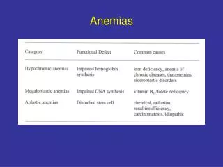
Macrocytic anemias • These can be divided into megaloblastic and non-megaloblastic types, depending on bone marrow findings. • MEGALOBLASTIC ANAEMIA • characterized by the presence in the bone marrow of erythroblasts with delayed nuclear maturation because of defective DNA synthesis (megaloblasts). • Megaloblasts are large and have large immature nuclei. • A characteristic abnormality of white cells, giant metamyelocytes, is frequently seen in megaloblastic anaemia.
Megaloblastic anemia Haematological values • The MCV is characteristically > 96 fL unless there is a coexisting cause of microcytosis when there may be a dimorphic picture. • The peripheral blood film shows macrocytes with hypersegmented polymorphs. • If severe, there may be leucopenia and thrombocytopenia.
Megaloblastic anemia Megaloblastic changes occur in: • Vitamin B12 deficiency or abnormal vitamin B12 metabolism • Folic acid deficiency or abnormal folate metabolism • Other defects of DNA synthesis, such as • Congenital enzyme deficiencies in DNA synthesis (e.g. oroticaciduria). • Acquired causes as therapy with drugs interfering with DNA synthesis (e.g. hydroxycarbamide (hydroxyurea), azathioprine, zidovudine - AZT) • Myelodysplasia due to dyserythropoiesis.
Biochemical basis of megaloblastic anemia • The key biochemical problem common to both vitamin B12 and folate deficiency is a block in DNA synthesis owing to an inability to methylate deoxyuridine monophosphate to deoxythymidine monophosphate, which is then used to build DNA. • The methyl group is supplied by the folatecoenzyme, methylene tetrahydrofolate. • Deficiency of folate reduces the supply of this coenzyme; • deficiency of vitamin B12 also reduces its supply by slowing the demethylation of methyltetrahydrofolate (methyl THF).
Vitamin B12 • Vitamin B12 is synthesized by certain microorganisms, and humans are ultimately dependent on animal sources. • It is found in meat, fish, eggs and milk, but not in plants. Vitamin B12 is not usually destroyed by cooking. • The average daily diet contains 5-30 μg of vitamin B12, of which 2-3 μg is absorbed. • The main function of B12 is the methylation of homocysteine to methionine with the demethylation of methyl THF polyglutamate to THF. THF is a substrate for folate polyglutamate synthesis
Vitamin B12 Absorption and transport • Vitamin B12 is liberated from protein complexes in food by gastric enzymes and then binds to a vitamin B12-binding protein ('R' binder) related to plasma transcobalamin I (TCI), derived from saliva. • Vitamin B12 bound to 'R' binder is released by pancreatic enzymes and becomes bound to intrinsic factor. • Intrinsic factor is a glycoprotein secreted by gastric parietal cells. It combines with vitamin B12 and carries it to specific receptors on the surface of the mucosa of the ileum.
Vitamin B12 enters the ileal cells and intrinsic factor remains in the lumen. • Vitamin B12 is transported from the enterocytes to the bone marrow and other tissues by the glycoprotein transcobalamin II (TCII).
Vitamin B12 Causes of vitamin B12 deficiency • Low dietary intake (vegans). • Impaired absorption ; - Stomach causes • Pernicious anaemia • Gastrectomy • Congenital deficiency of intrinsic factor - Small bowel causes • Ileal disease or resection • Malabsorption as Tropical sprue, celiac disease.
Pernicious anaemia • Pernicious anaemia (PA) is an autoimmune disorder in which there is atrophic gastritis with loss of parietal cells in the gastric mucosa with consequent failure of intrinsic factor production and vitamin B12 malabsorption.
Pathogenesis of Pernicious anaemia • This disease is common in the elderly, more common in females than males and those who have the blood group A. • There is an association with other autoimmune diseases, particularly thyroid disease, Addison's disease and vitiligo. • There is a higher incidence of gastric carcinoma with PA than in the general population. • Parietal cell antibodies are present in the serum in 90% of patients with PA. while intrinsic factor antibodies, found in only 50% of patients. • B12 deficiency may rarely occur in children from a congenital deficiency or abnormality of intrinsic factor, or as a result of early onset of the adult autoimmune type.
Pathology of Pernicious anaemia • Autoimmune gastritis affecting the fundus is present. • The parietal and chief cells are replaced by mucin-secreting cells • There is achlorhydria and absent secretion of intrinsic factor. • The histological abnormality can be improved by corticosteroid therapy, which supports an autoimmune basis for the disease.
Clinical features of Pernicious anaemia • The onset of PA is insidious, with progressively increasing symptoms of anaemia. • Jaundice (lemon-yellow colour owing to a combination of pallor and mild jaundice caused by excess breakdown of haemoglobin). • Glossitis (Red sore tounge) and angular stomatitis are sometimes present.
Neurological abnormalities • The neurological changes, if left untreated can be irreversible. • The classical neurological features are those • 1-Polyneuropathy involving peripheral nerves and the posterior and eventually the lateral columns of the spinal cord (subacute combined degeneration SCD);. Patients present with symmetrical paraesthesiae in the fingers and toes, early loss of vibration sense and proprioception, and progressive weakness and ataxia, Paraplegia may result.
2-Dementia, psychiatric problems, e.g. depression, hallucinations, delusions.
Investigations of Pernicious anaemia • CBC and blood film show the features of a megaloblastic anaemia . • Bone marrowshows the typical features of megaloblastic erythropoiesis (frequently not performed) • Serum bilirubin may be raised as a result of ineffective erythropoiesis where the destruction of developing red cells is much increased. • Serum vitamin B12 is usually low • Serum folate level is normal or high. • LDH: may be raised due to hemolysis. • Serum MMA& HC: raised.
Schilling test • Vitamin B12 absorption tests are performed only occasionally when the underlying cause of the B12 deficiency is not obvious. • Radioactive B12 is given orally followed by an i.m. injection of non-radioactive B12 to saturate B12 binding proteins and to flush out 58Co-B12. The urine is collected for 24 hours and > 10% of the oral dose would be excreted in a normal person. If this is abnormal, the test is repeated with the addition of oral intrinsic factor capsules. If the excretion is now normal, the diagnosis is pernicious anaemia or gastrectomy. If the excretion is still abnormal, the lesion must be in the terminal ileum or there may be bacterial overgrowth. The latter could be confirmed by repeating the test after a course of antibiotics.
Gastrointestinal investigations; - In PA there is achlorhydria. - Endoscopy or barium meal examination of the stomach is performed only if gastric symptoms are present.
Folic acid • Folic acid monoglutamate is not itself present in nature but occurs as polyglutamates. • Dietary intake: Folate is found in green vegetables such as spinach and broccoli, and offal, such as liver and kidney. Cooking causes a loss of 60-90% of the folate. The minimal daily requirement is about 100 μg. • There is no simple relationship between maternal folate status and fetal abnormalities but folic acid supplements at the time of conception and in the first 12 weeks of pregnancy reduce the incidence of neural tube defects.
Causes of folate deficiency • Nutritional (major cause) • Poor intakeas in Old age, Poor social conditions, Starvation, Alcohol excess (also causes impaired utilization) • Poor intake due to anorexia. • Gastrointestinal disease, e.g. partial gastrectomy, coeliac disease, Crohn's disease • Cancer. 3. Antifolatedrugsas Anticonvulsants (phenytoin, primidone), Methotrexate, Pyrimethamine, Trimethoprim.
Causes of folate deficiency • Excess utilization - Physiological as Pregnancy, Lactation, Prematurity - Pathological • Haematological disease with excess red cell production, e.g. haemolysis • Malignant disease with increased cell turnover • Inflammatory disease • Metabolic disease, e.g. homocystinuria • Haemodialysis or peritoneal dialysis • Malabsorption; Occurs in small bowel disease, but the effect is minor compared with that of anorexia
Clinical featuresof folate deficiency • Patients with folate deficiency may be asymptomatic or present with symptoms of anaemia or of the underlying cause. • Glossitis can occur. • Unlike with B12 deficiency, neuropathy does not occur
Investigationsof folate deficiency • CBC and blood film findings are those of a megaloblastic anaemia. • Blood measurements Serum and red cell folate are low. • Further investigations: when the cause is not obvious from the clinical picture or dietary history, Occult gastrointestinal disease should then be suspected and appropriate investigations, such as small bowel biopsy, should be performed.
Treatment and prevention of megaloblastic anaemia • Treatment depends on the type of deficiency. • Blood transfusion is not indicated in chronic anaemia. • Folic acid may produce a haematological response in vitamin B12 deficiency but may aggravate the neuropathy. • Large doses of folic acid alone should not be used to treat megaloblastic anaemia unless the serum vitamin B12 level is known to be normal. In severely ill patients, it may be necessary to treat with both folic acid and vitamin B12 while awaiting serum levels.
Treatment of vitamin B12 deficiency • Hydroxocobalamin 1000 μg can be given intramuscularly to a total of 5-6 mg over the course of 3 weeks; 1000 μg is then necessary every 3 months for the rest of the patient's life. • Alternatively, it is now recommended that oral B12 2mg per day is given, as 1-2% of an oral dose is absorbed by diffusion and therefore does not require intrinsic factor. • The use of sublingual nuggets of B12 (2 × 1000 μg daily) has been suggested to be an effective and more convenient option.
Treatment of vitamin B12 deficiency • Clinical improvement within 48 hrs,: • Reticulocytosis may occur within 2-3 days. • Improvement of the polyneuropathy may occur over 6-12 months, but long-standing spinal cord damage is irreversible. • Hypokalaemia can occur and, if severe, supplements should be given. • Iron deficiency often develops in the first few weeks of therapy. • Hyperuricaemia also occurs but clinical gout is uncommon.
Treatment of folate deficiency • Folate deficiency can be corrected by giving 5 mg of folic acid daily; • The same haematological response occurs as seen after treatment of vitamin B12 deficiency. • Treatment should be given for about 4 months to replace body stores. • Any underlying cause, e.g. coeliac disease, should be treated. • Prophylactic folic acid (400 μg daily) is recommended for all women planning a pregnancy. Many authorities also recommend prophylactic administration of folate throughout pregnancy. Prophylactic folic acid is also given in chronic haematological disorders where there is rapid cell turnover. A dose of 5 mg each week is probably sufficient.
MACROCYTOSIS WITHOUT MEGALOBLASTIC CHANGES • A raised MCV with macrocytosis on the peripheral blood film can occur with a normoblastic rather than a megaloblastic bone marrow. • A common physiological cause of macrocytosis is pregnancy and newborn.
MACROCYTOSIS WITHOUT MEGALOBLASTIC CHANGES • Common pathological causes are: • alcohol excess • liver disease • reticulocytosis • hypothyroidism • some haematological disorders (e.g. aplastic anaemia, sideroblastic anaemia, pure red cell aplasia) • drugs (e.g. cytotoxics - azathioprine) • spurious (agglutinated red cells measured on red cell counters) • cold agglutinins due to autoagglutination of red cells (the MCV decreases to normal with warming of the sample to 37°C).
In all these conditions, normal serum levels of vitamin B12 and folate will be found. The exact mechanisms in each case are uncertain, but in some there is increased lipid deposition in the red cell membran