Download
1 / 52
720 likes | 1.38k Vues
ORGANIZATION OF NERVOUS SYSTEM. DR. ZAHOOR ALI SHAIKH LECTURE--- 1. First We Will Discuss ‘ORGANIZATION & CELLS OF NERVOUS SYSTEM’ then we will talk about ‘Overview of Central nervous system’ [CNS]. ORGANIZATION OF NERVOUS SYSTEM. The Nervous system is organized into

E N D
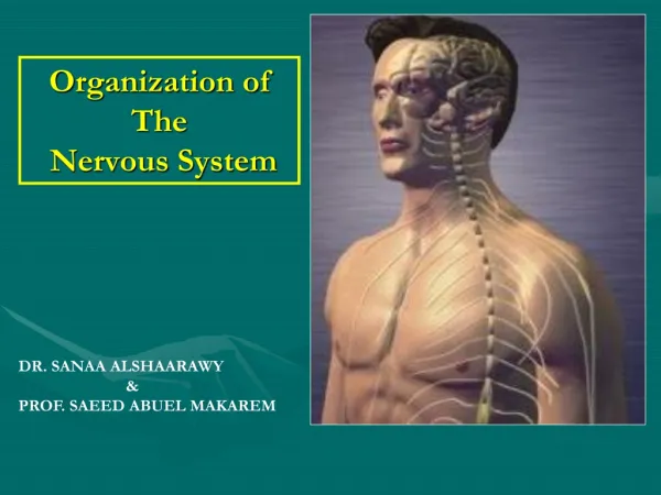
ORGANIZATION OF NERVOUS SYSTEM DR. ZAHOOR ALI SHAIKH LECTURE--- 1
First We Will Discuss ‘ORGANIZATION & CELLS OF NERVOUS SYSTEM’ then we will talk about ‘Overview of Central nervous system’ [CNS]
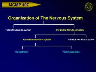
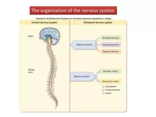
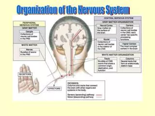
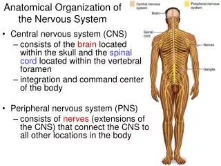
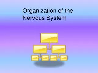
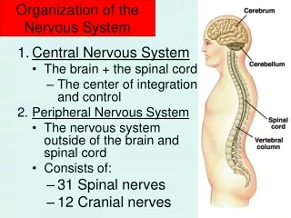
ORGANIZATION OF NERVOUS SYSTEM • The Nervous system is organized into 1. Central Nervous System (CNS) -- BRAIN -- SPINAL CORD 2. Peripheral Nervous System -- Nerve fibers that carry information between CNS and other parts of body at periphery.
CNS • Central nervous system regulates body activities. • CENTRAL NERVOUS SYSTEM 1) BRAIN 2) SPINAL CORD
PERIPHERAL NERVOUS SYSTEM PNS • Peripheral Nervous System is sub divided into 1) Afferent Division (Sensory) – which carry information to CNS 2) Efferent Division (Motor) – which carry information from CNS to muscle and glands.
PNS [cont] • Efferent (Motor) division of PNS is further divide into --Somatic Nervous System– it is under our voluntary control E.g. fibers of motor neuron that supply skeletal muscle. --Autonomic Nervous System – it is not under our control ( involuntary) and supply smooth muscle and glands.
PNS [cont] • Autonomic Nervous System (ANS) • ANS– supplies cardiac muscle, smooth - muscle, glands. ANS is divided into 1) Sympathetic ANS 2) Parasympathetic ANS • Enteric ANS– in the wall of digestive tract
PERIPHERAL NERVOUS SYSTEM • Neurons in peripheral nervous system transmit signals between the central nervous system and receptors ,and Effectors in the body. • In Peripheral Nervous System, there are 12 pairs of cranial nerves. • There are 31 pairs of spinal nerve, from spinal cord. - 31 pair of Spinal nerve exit from cervical (8 pairs), thoracic (12 pairs), lumber (5 pairs), sacral (5 pairs), and coccygeal (1 pair).
Cranial Nerves 12 pairs
Spinal Nerves 31 pairs
FUNCTIONAL CLASSES OF NEURONS • There are THREE functional types of Neurons 1) Afferent Neurons– which carry information to CNS. 2) Efferent Neurons– which carry information away from CNS (to the periphery). 3) Interneurons– They form interactive net-work between neuron.
AFFERENT NEURON • Afferent Neuron has sensory RECEPTOR , that generates action potential in response to a particular stimulus. Sensory impulse are taken by axon toward the spinal cord.
EFFERENT NEURON • Efferent Neuron lies in the peripheral nervous system. It has cell body in the CNS . Efferent axon leaves the cell body and goes to innervate muscle.
INTERNEURONS • Interneurons lie in the CNS. About 99% of all neurons are Interneurons. Human CNS has more than 100 billion Interneurons.
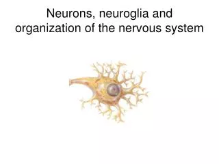
GLIAL CELLS or NEUROGLIA • Glial cells are connective tissue of CNS. • Glial cells support the interneurons physically, metabolically, and functionally. • They do not conduct nerve impulses. • Glial cells are of FOUR types.
GLIAL CELLS or NEUROGLIA (cont) • There are 4 type of Glial Cells 1. Astrocytes 2. Oligodendrocytes 3. Microglia 4. Ependymal Cells
Important Functions Of Glial Cells 1. Astrocytes • Functions i. Formation of blood – brain – barrier ii. Formation of neural scar tissue 2. Oligodendrocytes • Functions i. Forms myelin sheath in the CNS.
Important Functions Of Glial Cells 3. Microglia • Functions i. Phagocytosis [defense cells of CNS] ii. Release nerve growth factor. 4. Ependymal Cells • Functions i. Formation of Cerebrospinal fluid [CSF]. ii. Work as Neural Stem Cell – to form new neurons and glial cells.
Protection Of Brain • As CNS is very delicate, it is protected by – skull bone [covers the brain] and Vertebral column [surrounds the spinal cord]. • Meninges – cover brain and spinal cord. Meninges lie between bony covering [outside] and nervous tissue [inside]. Meninges are Durameter, Arachnoid and Piameter. • CSF - Cerebro Spinal Fluid is present in subarachnoid space. CSF works as cushion. • Blood - brain barrier – it selectively allows the materials to pass to brain.
Cerebro Spinal Fluid [CSF] • It surrounds brain and spinal cord. • It is present in subarachnoid space. • It is formed by choroid plexuses [capillaries in the piameter] of ventricles in the brain. • Volume of CSF is about 125 – 150 ml.
CSF [cont] • Clinical Note • Hydrocephalus [water in the brain]. • Occurs if CSF accumulates more, due to the block in its circulation or reabsorption. • If hydrocephalus is untreated, increased CSF pressure can lead to brain damage and mental retardation. • Treatment – surgically shunting the excess CSF to veins elsewhere in the body.
‘Important Information’ • Brain function depends on 1. Oxygen 2. Glucose • Brain needs continuous supply of O2 and Glucose. • Brain damage results if - brain gets no O2 supply for 4 to 5mins or - no glucose supply for 10 to 15mins.
OVER VIEW OF CNS • Parts of Brain (from top to bottom) • 1. Forebrain a) Cerebrum i) Cerebral cortex ii) Basal nuclei b) Diencephalon i) Thalamus ii) Hypothalamus
OVERVIEW OF CNS (cont) • Parts of Brain (cont) • 2. Brain stem - Mid brain - Pons - Medulla • 3. Cerebellum
CEREBRUM • Cerebrum constitutes 80% of total brain weight. • Outer layer, i.e. cerebral cortex of cerebrum is highly convoluted. • It has gyri [ridges] andsulci [depression].
Basal Nuclei • Basal Nuclei are present deep in the cerebrum. • Functions: - Co-ordination of movements - Muscle tone regulation
Diencephalon • Diencephalon is present in the interior of cerebrum. • It has 2 components: - Thalamus - Hypothalamus • Thalamus • All sensory information passes. • Crude awareness of sensation. • Hypothalamus • Regulates body temperature, has thirst and food intake center, regulates autonomic nervous system.
Brain Stem • Brain Stem [mid brain, Pons, Medulla]. • Brain Stem is continuous below with spinal cord. • Functions: • Majority of cranial nerves originate from brain stem. • Control Center for cardiovascular, respiratory system. • Regulation of postural reflexes. • Role in sleep – wake cycle.
Cerebellum • Cerebellum is attached at the back portion of brain stem. • Functions: • Balance of body. • Muscle tone. • Co-ordination and planning of skilled movements e.g. dance.
CEREBRUM • Cerebrum is the largest portion human brain. It is divided into TWO halves, Right and Left cerebral hemispheres. They are connected by CORPUS CALLOSUM which consists of about 300 million axons connecting two cerebral hemispheres. • Cerebral cortex– It is the outer shell of Gray matter covering the inner white matter.
CEREBRUM (cont) • Q—What is the GRAY Matter ? • A– It is the Cerebral cortex , which consists of cell bodies and their dendrites, as well as connective tissue glial cells. • Q– What is the WHITE Matter ? • A– It is the myelinated nerve fibers (Axons) . Its white appearance is due to Myelin sheath (lipid layer).
CEREBRAL CORTEX • NOTE – Gray matter of cerebral cortex is like computers of CNS. • White matter is like wires that connect the computers to each other.
CEREBRAL HEMISPHERE • Each cerebral hemisphere is divided into FOUR LOBES. • 1) Frontal lobe • 2) Parietal lobe • 3) Temporal lobe • 4) Occipital lobe
LOBES OF BRAIN • Central Sulcus separates the Frontal and Parietal lobe . • Frontal Lobe • It is located at the front and at the top. • It has MOTOR CORTEX area in the PRE -CENTRAL GYRUS- which controls the motor activity. • Motor speech area. • Elaboration of Thought.
LOBES OF BRAIN • Parietal lobe • It is located posterior to the central sulcus. • It has sensory cortex at post central gyrus. • Temporal Lobe • Located laterally [on the sides of head]. • It has auditory cortex.
LOBES OF BRAIN • Occipital Lobe • Located posteriorly [back of head]. • It has visual cortex ( center).