Download
1 / 1
10 likes | 115 Vues
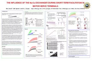
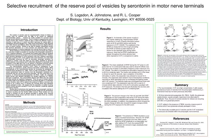
Selective recruitment of the reserve pool of vesicles by serontonin in motor nerve terminals S. Logsdon, A. Johnstone, and R. L. Cooper Dept. of Biology, Univ of Kentucky, Lexington, KY 40506-0025. Results. Introduction.

E N D
Selective recruitment of the reserve pool of vesicles by serontonin in motor nerve terminals S. Logsdon, A. Johnstone, and R. L. Cooper Dept. of Biology, Univ of Kentucky, Lexington, KY 40506-0025 Results Introduction The number of vesicles used for neurotransmitter release is limited, so vesicles must be retrieved from the NMJ to be reused for additional neurotransmitter release. Vesicles are readily available for recurrent fusion through several different mechanisms. There is evidence suggesting that when a vesicle recycles after release it might be able to follow two different routes for recycling: a rapid loop, or a slower one which reprocess the vesicle within an endoplasmic reticulum (Klingauf et al., 1998; Kuromi and Kidokoro, 2002; Richards et al., 2000; Stevens and Williams, 2000). This might produce two slightly different pools of reserve vesicles. Results in the field of synaptic transmission predict that electrical stimulation of a nerve terminal can selectively utilize a rapid recycling population of vesicles separately from vesicles that are kept in reserve. Recent findings of vesicular kinetics and recycling processes also support the notion that it might be possible for recycling in the presynaptic membrane to occur in different pathways. It is possible that multiple paths might be regulated independently by second messengers, such as those activated by 5-HT. However, direct electrical stimulation of nerve terminals could use a different intracellular path than is used for 5-HT. In the crayfish motor nerve terminals, one route might be differentially influenced by 5-HT via secondary messenger activation, as compared to another route that might be more tightly regulated by electrical activity patterns of the terminal. Such a differential regulation is reasonable since incubation of the NMJ with 5-HT in the absence of persistent electrical activity results in more vesicles being released with a single or within a train of stimuli. The neuromodulator serotonin (5-HT), which is endogenous to the hemolymph of crustaceans, greatly enhances transmitter release (Dudel, 1965a) presynaptically at the neuromuscular junction by increasing the probability of vesicular fusion (Southard et al., 2000). 5-HT, at a high concentration (1 μM), also causes spontaneous fusion of vesicles from the presynaptic membrane by a mechanism that has not been previously described. We have noted that in the presence of dl-threo-beta-benzyloxyaspartate (DL-TBOA, 10μM), the glutamate uptake blocker, with repeated stimulation the synaptic responses (i.e., EPSPs) attenuate in amplitude. The evoked EPSPs depress with TBOA since the rapidly recycling vesicles are depleted of glutamate. Thus, the pool of evoked vesicles for release might not be depleted, but they are recycling with little or no glutamate present. However, when 5-HT is added in the presence of TBOA, a new pool, also referred to as a reserve pool, of vesicles are recruited which have glutamate already packaged in them. We suggest that the population of readily recycling vesicles during electrical activation of the nerve terminal is distinctly separate from reserve vesicles. Studies about mechanisms that regulate the use of presynaptic vesicles in motor nerve terminals at chemical synapses provide insight into the mechanisms of vesicle recycling and the kinetics within neurons. These studies are very relevant to humans as well as general principals in biology since chemical communication among nerves and from nerves to muscles work by a similar means in all living animals. Figure 1: A schematic of the opener muscle in the crayfish walking leg. Representative EPSP responses to a train of twenty stimulation pulses given at 40 Hz and 60Hz before and during exposure to 5-HT (100nM). The amplitude of the EPSPs is measured from the trough preceding the EPSP of interest to peak response, as shown for the twentieth pulse during saline exposure. Note that the EPSP amplitudes are greater throughout the entire stimulus train when exposed to 5-HT in comparison to saline. Figure 5: A schematic representation of the vesicle pathways within the presynaptic motor nerve terminal. (A) In the absence of electrical stimulation (i.e., Inactive, A) of the nerve terminal few vesicles will spontaneously be released at slow rate; thus, a slow recycling path (path 1 to 2) will be utilized. Upon electrical stimulation (i.e., Active, B) the high-output synapse (Sy1), the one with two active zones as shown with dark hemispheres sitting on the inner face of the synapse, will be recruited before the low-output synapse (Sy2) with only one active zone (B). Vesicles maybe recruited from the reserve vesicle pool (RV, path 1) to the readily releasable vesicle pool (RRV) for docking and fusion with the presynaptic membrane as well during electrical stimulation. The vesicles may then recycle through a slow process and intermediate endosomal (ES) stage (path 2), as well as a different path (path 3) that is relatively rapid in recharging the vesicles with transmitter before the vesicle ends up back at the RRV pool. When the nerve terminal is not electrical stimulated but is exposed to 5-HT (i.e., 5-HT & Inactive, C), at a low concentration (100nM), there is a priming of the synapses by promoting path 1 (C). The activation of the second messenger IP3 can recruit vesicles from RV as well as enhance priming and docking of vesicles at the synapses within the RRV by phosphorylating synaptically relevant proteins. In this case, both the high-output (Sy1) and low-output (Sy2) synapses will be influenced. When combining electrical activity and exposure to 5-HT (i.e., 5-HT & Active, D) a marked enhancement of transmission will occur which will activate path 1 and path 3 for rapid recycling. Figure 2: The mean amplitude of EPSP during the 10th pulse in a 20 pulse train for 10 consecutive trials was used to show the effects over time due to the increase in stimulation frequency and/or exposure of 5-HT (100nM). The effect of increasing the stimulation frequency from 40 to 60 Hz is relatively rapid. The means and standard error of the mean is shown for each 50 seconds. Upon completion of the 60Hz stimulation in saline, the preparation was allowed to recover for 5 minutes. The responses were tested to insure that baseline conditions resumed for 40Hz stimulation prior to exchanging the bathing media with 5-HT solution. The bathing media was exchanged three times with one containing 5-HT. The preparation was then left to soak for 5 minutes before data collection in the 40 Hz stimulation paradigm with 5-HT exposure. The break in the time axis illustrates this recovery and incubation time before adding the 5-HT containing bath. Summary 1.The neuromodulator, 5-HT, at a high concentration (1 μM) causes spontaneous fusion of vesicles from the presynaptic membrane by a mechanism that has not been previously described. 2. Dl-threo-beta-benzyloxyaspartate (DL-TBOA, 10μM), the glutamate uptake blocker, causes synaptic responses to be attenuated in amplitude. Thus, the pool of evoked vesicles for releaseare recycling with little or no glutamate present. 3. 5-HT, added in the presence of TBOA, recruits a reserve pool of vesicles which have glutamate already packaged in them. 4. The electrically excitable pool of vesicles and the 5-HT modulated vesicle pool are divisible within the presynaptic nerve terminal. Figure 3: The percent change in the 10th (A) and 20th (B) EPSP amplitudes for each preparation induced by 5-HT during the 40Hz and 60Hz stimulation indicated that a smaller change occurred for the 60Hz paradigm. In addition, the preparations which showed the largest change at 40Hz also showed the largest change at 60Hz during the 5-HT exposure for the 20th event. Methods Animals Mid-sized crayfish (Procambarus clarkii), measuring 8 - 10 cm in body length and weighing 20 to 36 grams, were obtained from Atchafalaya Biological Supply Co. (Raceland, LA). Animals were housed in an aquatic facility within the laboratory in individual tanks, and were fed fish food pellets every three days. Only male crayfish in their intermolt stage were used. Dissection & Physiology In brief, the experimental paradigm is to record intracellular EPSPs from muscle fibers during stimulation of the motor nerve in the presence of saline and then saline containing TBOA. As the EPSP responses get smaller and unable to be discerned, a saline containing 5-HT is used. The second phase of experiments consist of using a similar experimental paradigm but to record quantal responses by use of a focal macropatch electrode placed over a defined region of the motor nerve terminal. Excitatory Postsynaptic Potentials (EPSPs) in crayfish: EPSPs at the crayfish NMJ were recorded by intracellular electrodes, with 30‑60 MΩ resistance microelectrodes, filled with 3 M KCl. Responses were recorded with a standard intracellular electrode amplifier (AxoClamp 2A, Axon Instruments). Electrical signals were recorded onto VHS tape and on‑line to a Power Mac 9500 via a MacLab/4s interface. EPSPs were recorded at 10 kHz. All events were appropriately scaled to known values measured on an oscilloscope. The opener muscle preparations were stimulated to induce a short-term facilitation (STF) by giving a 40 Hz train of ten pulses at intervals of 5 or 10 seconds. Figure 4: The presence of TBOA resulted in a run down of evoked transmission. Here the 10th EPSP amplitude in measured over time. After a short while the EPSP amplitude is not detectable, but after adding 5-HT (1um) the evoked EPSP responses are revived to larger amplitudes than control levels. References Southard RC, Haggard J, Crider ME, Whiteheart SW, and Cooper RL. 2000. Influence of serotonin on the kinetics of vesicular release. Brain Research 871:16‑28. Sparks G and Cooper RL. 2004. 5-HT offsets homeostasis of synaptic transmission during short-term facilitation. (In Press- J. of Applied Physiology). Tabor Jand Cooper RL. 2002. Physiologically identified 5-HT2 -like receptors at the crayfish neuromuscular junction. Brain Research 932:91-98. Funding was provided by NSF grant IBN-0131459 (RLC) and a G. Ribble Fellowship for undergraduate studies in the Department of Biology at the University of Kentucky (SL). 2mV