Download
1 / 1
10 likes | 221 Vues
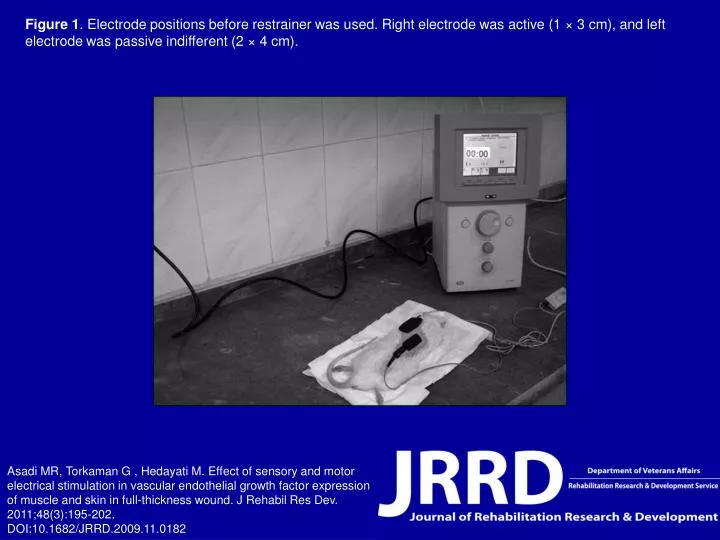
Figure 1 . Electrode positions before restrainer was used. Right electrode was active (1 × 3 cm), and left electrode was passive indifferent (2 × 4 cm).

E N D
Figure 1. Electrode positions before restrainer was used. Right electrode was active (1 × 3 cm), and left electrode was passive indifferent (2 × 4 cm). Asadi MR, Torkaman G , Hedayati M. Effect of sensory and motor electrical stimulation in vascular endothelial growth factor expression of muscle and skin in full-thickness wound. J Rehabil Res Dev. 2011;48(3):195-202.DOI:10.1682/JRRD.2009.11.0182