Download
1 / 3
30 likes | 157 Vues
SORF2. SORF3. US10. US1. US10. EGFP. A. 1 2 3. B. 23 kb. 9.4 kb. C. 6.6 kb. US10-EGFP. EGFP. Mock. 4.4 kb. 52 KD. 2 kb. 28 KD. 1.5 kb.

E N D
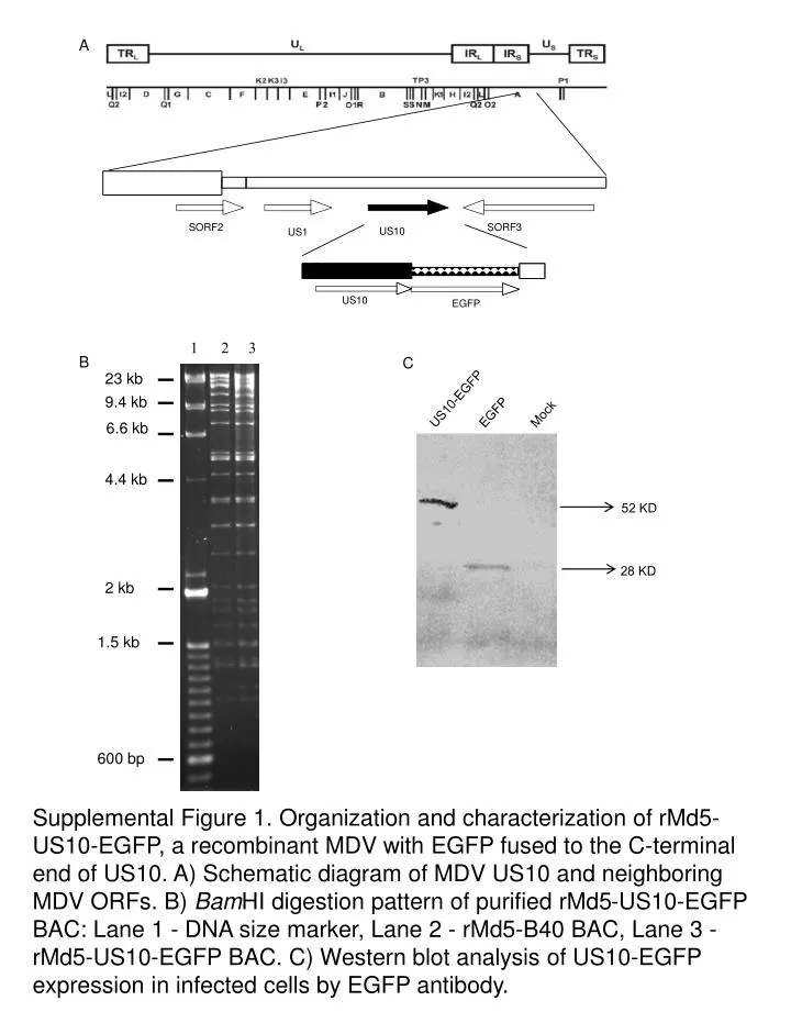
SORF2 SORF3 US10 US1 US10 EGFP A 1 2 3 B 23 kb 9.4 kb C 6.6 kb US10-EGFP EGFP Mock 4.4 kb 52 KD 2 kb 28 KD 1.5 kb Supplemental Figure 1. Organization and characterization of rMd5-US10-EGFP, a recombinant MDV with EGFP fused to the C-terminal end of US10. A) Schematic diagram of MDV US10 and neighboring MDV ORFs. B) BamHI digestion pattern of purified rMd5-US10-EGFP BAC: Lane 1 - DNA size marker, Lane 2 - rMd5-B40 BAC, Lane 3 - rMd5-US10-EGFP BAC. C) Western blot analysis of US10-EGFP expression in infected cells by EGFP antibody. 600 bp
A B No. of Plaques log10 Days post infection Supplemental Figure 2. Flow cytometry analysis of CEF infected with the rMd5-US10-EGFP virus and growth curve analysis and western blot analysis of US10-EGFP. A) EGFP expression in virus-infected cells was analyzed at 3 dpi (green line) and 5 dpi (blue line) with flow cytometry; the black line is the uninfected control CEF. B) rMd5-B40 and rMd5-US10-EGFP growth curves; rMd5-B40 and rMd5-US10-EGFP are indicated by triangles and squares, respectively.
A B C Supplemental Figure 3. Subcellular localization of EGFP in rMd5-EGFP virus-infected cell.The recombinant MDV with EGFP replacing US10 was used to infect CEF, then visualized using confocal microscopy at 3 dpi. A) rMd5-EGFP virus-infected CEF culture with Hoechst 33342 staining. B) EGFP was visualized through fluorescence. C) Merged image of A and B.