Download
1 / 52
880 likes | 2.14k Vues
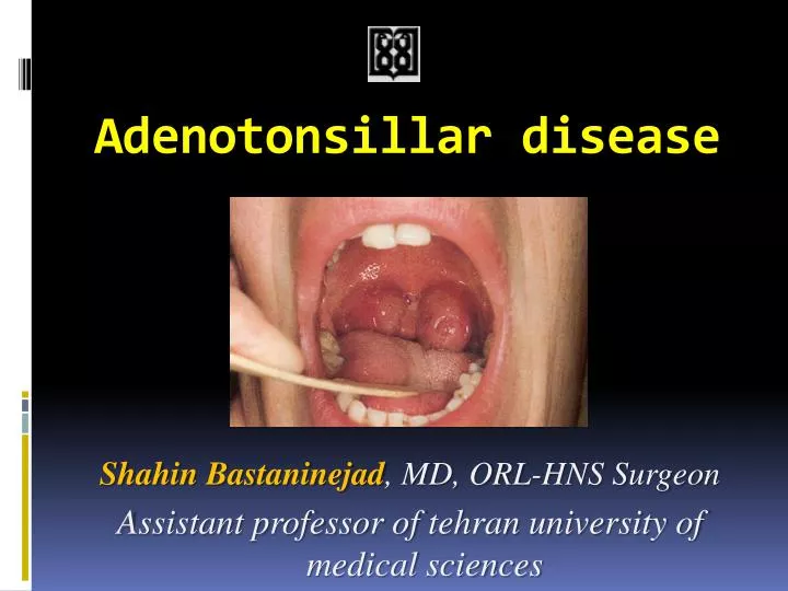
Adenotonsillar disease. Shahin Bastaninejad , MD, ORL-HNS Surgeon Assistant professor of tehran university of medical sciences. Anatomy. Tonsil boundary Plica triangularis Adenoid boundary Posterior aspect of the nasal septum Fossa of Rosenmüller Passavant’s ridge. Waldeyer’s Ring.

E N D
Adenotonsillar disease ShahinBastaninejad, MD, ORL-HNS Surgeon Assistant professor of tehran university of medical sciences
Anatomy Tonsil boundary • Plicatriangularis Adenoid boundary • Posterior aspect of the nasal septum • Fossa of Rosenmüller • Passavant’s ridge
Presentation outlines • Acute Infections • Chronic diseases • Obstructive hyperplasia • Mass • Surgery
Acute Adenotonsillitis Etiology • 85% of this problem is due to the viral infection (less in children) • In bacterial infections there is about 40% antibiotic resistancy (due to beta-lactamase-producing germs) • GABHS is the most important pathogen because of potential sequelae
Bacteriology of adenotonsillitis • Group A beta-hemolytic is most recognized pathogen • This organism is associated with a risk of rheumatic fever and glomerulonephritis • Many other organisms are involved : • H.influenza • S. aureus • Streptococcus pneumoniae
GABHS • More common in 5 to 15 years old children • Not seen in less than 3 years
Diagnosis • Viral pharyngitis symptoms: • Coryza • Hoarseness • Cough • Conjunctivitis • Centor criteria for GABHS: • Hx of fever more than 38 • Anterior cervical LAP • Pharyngeal or Tonsillar exudate • Absence of cough
Approach to the Centor scoring • 0-1 Abx not needed • 2-4 perform Cx • Clue : when all 4 scores are present in 44% of the patients there is no GABHS
Treatment Plan • Delay in treatment up to 9 days can be acceptebale • When empiric txy? • Lack of Pt .f/u • Lack of Lab. access • Toxic presentation • In some extends when all 4 measures present
In parentheses!!! • When culture is positive there are two possibilites: • True infection • Carrier state • In this scenario, serological evaluation with ASO(anti-streptolysin O) will be usefull (in true infection it will be more than 3 times than its usual range)
Medical Management • Penicillin is first line treatment oral medication is preferable (penicillin V) • Other choices: • Amoxicillin (wide spectrum than Pencillin V) • Macrolides • Clindamycin
Recurrent or unresponsive infections require treatment with beta-lactamase resistant antibiotics such as • Clindamycin • Augmentin • Penicillin plus rifampin (or Erythro + Metro)
If no response after 48 hr, re-evaluate patient for the followings: • Sequelea • Patient’s incompliance • Other underlying disease • Abx failure
Peritonsillar abscess • Abscess formation outside tonsillar capsule • Signs and symptoms: • Fever • Sore throat • Dysphagia/odynophagia • Drooling • Trismus • Unilateral swelling of soft palate/pharynx with uvula deviation
Peritonsillarabscess… • Thought to be extension of tonsillitis to involve surrounding tissue with abscess formation • Recently described to be an infection of small salivary glands in the supratonsillar fossa called Weber’s glands • Would explain superior pole involvement and the usual absence of tonsillar erythema/exudates
IMN • Clinical diagnosis can be made from the characteristic triad of fever, pharyngitis, and lymphadenopathy lasting for 1 to 4 weeks • Laboratory tests are needed for confirmation • Serologic test results include a normal to moderately elevated white blood cell count, an increased total number of lymphocytes (more than 50%), greater than 10% atypical lymphocytes, and a positive reaction to a "mono spot" test
IMN • When "mono spot" or heterophile test results are negative, additional laboratory testing may be needed to differentiate EBV infections from a mononucleosis-like illness • EBV-Specific Laboratory Tests: • IgM and IgG to the viral capsid antigen • IgM to the early antigen • antibody to EBNA
IMN – Test interpretation • Primary Infection: Primary EBV infection is indicated if IgM antibody to the viral capsid antigen is present and antibody to EBNA is absent • Past Infection: If antibodies to both the viral capsid antigen and EBNA are present, then past infection (from 4 to 6 months to years earlier) is indicated
IMN – Test interpretation • Reactivation: In the presence of antibodies to EBNA, an elevation of antibodies to early antigen suggests reactivation • Chronic EBV Infection: Reliable laboratory evidence for continued active EBV infection is very seldom found in patients who have been ill for more than 4 months
Chronic Tonsillitis • Chronic sore throat • Malodorous breath • Presence of tonsilliths • Persistent tender cervical lymphadenopathy • Lasting at least 3 months • Be aware of Anaerobic infections
Cryptic tonsils • Hyperkeratosis, mycosis leptothrica • Tonsilloliths
Obstructive Adenoid Hyperplasia • Signs and Symptoms • Obligate mouth breathing • Hyponasal voice • Snoring and other signs of sleep disturbance
Obstructive Tonsillar Hyperplasia • Snoring and other symptoms of sleep disturbance • Muffled voice • Dysphagia
Malignant Neoplasms • Most common is lymphoma • Non-Hodgkin’s lymphoma • Rapid unilateral tonsillar enlargement associated with cervical lymphadenopathy and systemic symptoms
Congenital tonsillar masses • Teratoma • Hemangioma • Lymphangioma • Cystic hygroma
Tonsillectomy(2010-AAOHNS) • Infection indications: • Pharyngitis more than 7 / yr in 1 yr • More than 5 / yr for 2yrs • More than 3 / yr for 3yrs • Recurrent infections with modifying factors: • Multiple Abx allergy / intolerance • PF.ASP.A: periodic fever/aphthous stomatitis and pharyngitis/adenitis • History of peritonsillar abscess
TnosillectomyCont… • Persistent foul taste or breath due to chronic tonsillitis not responsive to medical therapy • Chronic or recurrent tonsillitis associated with streptococcal carrier state and not responding to beta-lactamase resistant antibiotics • Unilateral tonsil hypertrophy presumed to be neoplastic
Adenotonsillectomy • ATH and Sleep disordered breathing (SDB) • Severity of the SDB depends on adenotonsillar size and/or Craniofacial anatomy and/or neuromuscular tone • Ask for comorbid conditions: Growth retardation / poor school performance / enuresis / behavioral problems (ADHD,…) • Polysomnography indications (PaO2 less than 85% and/or AHI>5) check PSG in obese patient/down syndrome/craniofacial anomaly &…
Adenoidectomy • Infection: • Purulent adenoiditis • Adenoid hypertrophy associated with: • Chronic otitis media with effusion • Chronic recurrent acute otitis media • Chronic otitis media with perforation • Otorrhea or chronic tube otorrhea • Obstruction (next slide) • Other: • Suspected neoplasia • Adenoid hypertrophy associated with chronic sinusitis
Adenoidectomy Cont… • Obstruction: • Adenoid hypertrophy associated with excessive snoring and chronic mouth-breathing • Sleep apnea or sleep disturbances • Adenoid hypertrophy associated with: • Corpulmonale • Failure to thrive • Dysphagia • Speech abnormalities • Craniofacial growth abnormalities • Occlusion abnormalities • Speech abnormalities
Pre-Op Evaluation ofAdenoid Disease • Triad of hyponasality, snoring, and mouth breathing • Rhinorrhea, nocturnal cough, post nasal drip • “Adenoid facies” • long face, crowded incisors
Pre-Op Evaluation of AdenoidDisease Evaluate palate • Symptoms/FH of CP or VPI • Bifid uvula • CNS or neuromuscular disease • Preexisting speech disorder?
Pre-Op Evaluation of Adenoid Disease Lateral neck films are useful only when history and physical exam are not in agreement. Accuracy of lateral neck films is dependent on proper positioning and patientcooperation.