Download
1 / 39
700 likes | 2.08k Vues
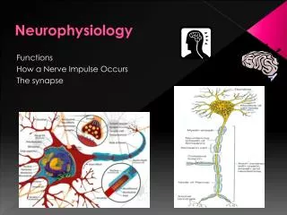
Neurophysiology. The Neuron: Pumps, Channels, and Membrane Potentials . Neuron. The functional and structural unit of the nervous system There are many, many different types of neurons but most have certain structural and functional characteristics in common. COMMUNICATION MODEL. Sender.

E N D
Neurophysiology The Neuron: Pumps, Channels, and Membrane Potentials
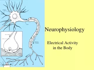
Neuron • The functional and structural unit of the nervous system • There are many, many different types of neurons but most have certain structural and functional characteristics in common
COMMUNICATION MODEL Sender Receiver Medium: AXON Message: ACTION POTENTIAL Function • Neurons are excitable cells (responsive to stimuli) specialized to conduct information (communicate) from one part of the body to another via electrical impulses (Action Potentials) conducted along the length of axons
_ _ _ _ _ _ _ _ _ _ _ _ _ _ _ _ _ _ _ _ _ _ _ _ _ _ _ _ _ _ _ Electric Current Electricity: flow of electrons through conductor e- e- e- e- e- e- e- e- e-e- e- e- e- e- e- e- e- Nerve impulse: flow of ions across membrane + + + + + + + + + + + + + +
Basic Concepts Review • Ions – charged particles • Anions – Negatively charged particles • Cations – Positively charged particles • Electrostatic forces • Opposite charges attract, same charges repel • Ions flow along their electrical gradient when they move toward an area of opposite charge • Concentration forces • Diffusion – movement of ions through semipermeable membrane • Ions flow along their chemical gradient when they move from an area of high concentration to an area of low concentration • Together, the electrical and chemical gradients constitute the Electrochemical gradient
Passive transport. Substances diffuse spontaneously down their concentration gradients, crossing a membrane with no expenditure of energy by the cell. The rate of diffusion can be greatly increased by transport proteins in the membrane. Active transport. Some transport proteins act as pumps, moving substances across a membrane against their concentration gradients. Energy for this work is usually supplied by ATP. ATP Diffusion. Hydrophobic molecules and (at a slow rate) very small uncharged polar molecules can diffuse through the lipid bilayer. Facilitated diffusion. Many hydrophilic substances diffuse through membranes with the assistance of transport proteins, either channel or carrier proteins. Review: Passive and active transport compared
Na+ binding stimulates phosphorylation by ATP. Cytoplasmic Na+ binds to the sodium-potassium pump. [Na+] high [K+] low 2 4 6 5 1 Na+ Na+ Na+ Na+ Na+ [Na+] low [K+] high ATP P Na+ CYTOPLASM ADP Na+ Na+ Na+ Phosphorylation causes the protein to change its conformation, expelling Na+ to the outside. K+ is released and Na+ sites are receptive again; the cycle repeats. 3 K+ P K+ K+ Loss of the phosphate restores the protein’s original conformation. Extracellular K+ binds to the protein, triggering release of the Phosphate group. K+ K+ K+ P P i Sodium-Potassium Pump • Is one type of active transport system • It is an electrogenic pump that generates the voltage across a membrane
Ion Channels – Membrane Potential • Membrane potential is the voltage difference across a membrane • Resting potential (when the cell is not firing) is a 70mV difference between the inside and the outside - the membrane is polarized • When gated ion channels open, ions diffuse across the membrane following their electrochemical gradients. • This movement of charge is an electrical current and can create voltage (measure of potential) energy change across the membrane. • This electrical charge across the membrane is the membrane potential.
Nernst/GHK Equations Predicts Membrane Potentials • Membrane potential is influenced by concentration gradient of ions and membrane permeability to those ions • A simplified equation at room temperature: • GHK equation predicts membrane potential using multiple ions
Resting Membrane Potential • The resting potential exists because ions are concentrated on different sides of the membrane: • Na+ and Cl- outside the cell • K+ and organic anions inside the cell • Due to different membrane permeabilities of the passive ion channels and operation of the sodium-potassium pump
Electrical Signals: Ion Movement • Resting membrane potential determined by • Na+ and K+ concentration gradients • Cell’s resting permeability to K+, Na+, and Cl– • Gated channels control ion permeability • Mechanically gated • Chemical gated • Voltage gated • Threshold varies from one channel type to another
Membrane Potentials: Signals • Neurons use changes in membrane potential to receive, integrate, and send information • Two types of signals are produced by a change in membrane potential: • Gradedpotentials (short-distance) • Action potentials (long-distance)
Signals Carried by Neurons • Resting neuron – membrane is polarized, inner, cytoplasmic side is negatively charged • Stimulation of the neuron depolarization • Strong stimulus applied to the axon triggers nerve impulse/action potential • Membrane becomes negative externally • Impulse travels the length of the axon • Membrane repolarizes itself
Signals Carried by Neurons Figure 12.9c–d
Graded Potentials • Short-lived, local changes in membrane potential • Currents decrease in magnitude with distance • Their magnitude varies directly with the strength of the stimulus – the stronger the stimulus the more the voltage changes and the farther the current goes • Sufficiently strong graded potentials can initiate action potentials
Action Potentials • Supra-threshold stimuli cause voltage-gated Na+ channels to open • Na+ to enters the cell down its electrochemical gradient to produce depolarizing currents that are translated into action potentials • Threshold Voltage– membrane is depolarized by ~ 15 mV stimulus • The AP is a brief reversal of membrane potential with a total amplitude of 100 mV (from -70mV to +30mV) • APs do not decrease in strength with distance All-or-None phenomenon – action potentials either happen completely, or not at all
Depolarization Phase • Na+ activation gates open quickly and Na+ enters causing local depolarization which opens more activation gates and cell interior becomes progressively less negative. • Threshold – a critical level of membrane potential (~ -55 mV) where depolarization becomes self-generating
Repolarization Phase • Positive intracellular charge reduces the driving force of Na+ to zero. Sodium inactivation gates of Na+ channels close. • After depolarization, the slower voltage-gated K+ channels open and K+ rapidly leaves the cell following its electrochemical gradient restoring resting membrane potential
Hyperpolarization • The slow K+ gates remain open longer than needed to restore the resting state. • This excessive efflux causes hyperpolarization of the membrane • The neuron is insensitive to stimulus and depolarization during this time
Propagation of an Action Potential • The action potential is self-propagating and moves away from the stimulus (point of origin)
Role of the Sodium-Potassium Pump • Repolarization restores the resting electrical conditions of the neuron, but does not restore the resting ionic conditions • Ionic redistribution is accomplished by the sodium-potassium pump following repolarization
Refractory Periods • Absolute refractory period is the time from the opening of the Na+ activation gates until the closing of inactivation gates, the neuron cannot respond to another stimulus • Relative refractory period follows the absolute refractory period. Na+ gates are closed, K+ gates are open and repolarization is occurring. Only a strong stimulus can generate an AP
Axon Conduction Velocities • Conduction velocities vary widely among neurons, determined mainly by: • Axon Diameter – the larger the diameter, the faster the impulse (less resistance) • Presence of a Myelin Sheath – myelination increases impulse speed (Continuous vs. Saltatory Conduction)
Myelin Sheath • A Schwann cell envelopes and encloses the axon with its plasma membrane. • The concentric layers of membrane wrapped around the axon are the myelin sheath • Neurilemma – cytoplasm and exposed membrane of a Schwann cell
Saltatory Conduction • Gaps in the myelin sheath between adjacent Schwann cells are called nodes of Ranvier (neurofibral nodes) • Voltage-gated Na+ channels are concentrated at these nodes • Action potentials are triggered only at the nodes and jump from one node to the next • Much faster than conduction along unmyelinated axons
Synapse • As the impulse reaches the axon terminals the signal is relayed to target cells at specialized junctions known as synapses • Synapse is a junction that mediates information transfer from one neuron to another neuron or to an effector cell
Synaptic Cleft: Information Transfer • Nerve impulses reach the axon terminal of the presynaptic neuron and open Ca2+ channels • Neurotransmitter is released into the synaptic cleft via exocytosis • Neurotransmitter crosses the synaptic cleft and binds to receptors on the postsynaptic neuron • Postsynaptic membrane permeability changes due to opening of ion channels, causing an excitatory or inhibitory effect
Synaptic Transmission • An AP reaches the axon terminal of the presynaptic cell and causes V-gated Ca2+ channels to open. • Ca2+ rushes in, binds to regulatory proteins & initiates NT exocytosis. • NTs diffuse across the synaptic cleft and then bind to receptors on the postsynaptic membrane and initiate some sort of response on the postsynaptic cell.
Neurotransmitter Removal • NTs are removed from the synaptic cleft via: • Enzymatic degradation • Diffusion • Reuptake
Effects of the Neurotransmitter • Different neurons can contain different NTs. • Different postsynaptic cells may contain different receptors. • Thus, the effects of an NT can vary. • Some NTs cause cation channels to open, which results in a graded depolarization. • Some NTs cause anion channels to open, which results in a graded hyperpolarization.
EPSPs & IPSPs • Typically, a single synaptic interaction will not create a graded depolarization strong enough to migrate to the axon hillock and induce the firing of an AP • However, a graded depolarization will bring the membrane potential closer to threshold. Thus, it’s often referred to as an excitatory postsynaptic potential or EPSP. • Graded hyperpolarizations bring the membrane potential farther away from threshold and thus are referred to as inhibitory postsynaptic potentials or IPSPs.
Excitatory And Inhibitory Neurotransmitters • If a transmitter depolarizes the post-synaptic neuron, it is said to be excitatory • If a transmitter hyperpolarizes the post-synaptic neuron, it is said to be inhibitory • Whether a transmitter is excitatory or inhibitory depends on its receptor
Excitatory And Inhibitory Neurotransmitters • Acetylcholine is excitatory because its receptor is a ligand-gated Na+ channel • GABA is inhibitory because its receptor is a ligand-gated Cl- channel • Other transmitters (e.g. vasopressin, dopamine) have G-protein-linked receptors • Effects depend on the signal transduction pathway and cell type Fig 48.7
Summation • One EPSP is usually not strong enough to cause an AP. However, EPSPs may be summed. • Temporal summation - the same presynaptic neuron stimulates the postsynaptic neuron multiple times in a brief period. The depolarization resulting from the combination of all the EPSPs may cause an AP • Spatial summation - multiple neurons all stimulate a postsynaptic neuron resulting in a combination of EPSPs which may yield an AP
Synaptic Organization • Communication between neurons is not typically a one-to-one event. • Sometimes a single neuron branches and its collaterals synapse on multiple target neurons. This is known as divergence. • A single postsynaptic neuron may have synapses with as many as 10,000 postsynaptic neurons. This is convergence.