Download
1 / 44
440 likes | 466 Vues
Pathophysiology of asthma and chronic obstructive pulmonary disease. M. Tatár. OBSTRUCTIVE LUNG DISEASES. localized : laryngeal constriction , tracheal and bronchial carcinoma , foreign bodies. generalized: asthma, COPD, bronchiectasis, cystic fibrosis.

E N D
Pathophysiology of asthmaand chronic obstructive pulmonary disease M. Tatár
OBSTRUCTIVE LUNG DISEASES localized: laryngealconstriction, tracheal and bronchialcarcinoma, foreignbodies generalized: asthma, COPD, bronchiectasis, cystic fibrosis OBSTRUCTIVE VENTILATORY DISORDER - spirometry Airflow limitation
End of quiet expiration - 0.5 - 2.5 0.5 Inspiration 0 0 0 0 0
Inspiration - 2.5 + 2.0 Forced expiration 0.5 - 2.0 - 1.5 - 1.0 - 0.5 0
Forced expiration + 2.0 0.5 + 2.5 + 1.0 0 + 2.0 + 1.5 EPP
ASTHMA - definition Heterogeneous, multifactorial disease with variable and mostly reversible respiratory pathway obstruction based on a chronic bronchial inflammatory reaction. Symptoms (wheezing, cough particularly at night and early morning, dyspnoea – chest tightness or shortness of breath) are variable and correlated with expiratory flow limitation. Bronchial hyperresponsiveness is often present
Volume Normal subject Asthmatic (after bronchodilator) Asthmatic (before bronchodilator) FEV1 1 2 3 4 5 Time (seconds)
Classification A. Allergicasthma • atopy, geneticpredisposition • IgE, mastcells and eosinophilsresponse to allergens A. Nonallergicasthma • no environmentalcausescanbeidentified • negative skin test to commonairbornallergens • rathernegativefamilyhistory C. Occupationalasthma • sensibilisation of airways to inhalantchemicals
Development of asthma Risk factors Predisposing - atopy, gender Causal - allergens, aspirin, chemicals Contributing - respiratory infections, diet, air pollution, smoking Factors that exacerbate asthma - triggers allergens, respiratory infections, exercise, emotions
Triggers – mechanisms of action Respiratoryinfections • epithelialdamage • airwayinflammation Exercise • hyperventilation (reflex airflowlimitation) - cooling of mucosa - osmolaritychanges of fluid liningepithelium Emotions (laughing, crying, anger, fear) • hyperventilation • hypocapnia (bronchioloconstriction)
Bronchial hyperresponsiveness Instability of the airways = exaggerated bronchoconstrictor response to a wide variety of stimuli Key factor - airway inflammation Mechanisms - direct and indirect
Airway hyperresponsiveness Direct antagonists (methacholine) Nerve Airway with limited airflow SO2, bradykinin Mediators Indirect agonists (exercise, hypotonic or hypertonic aerosols Mast cell
antihyperreactivefactors prohyperreactive factors 2-adrenergic -adrenergic VIP/PHM cholinergic anticholinergic SP/NK neutral peptidases free radicals antioxidants peptidases (Neu) corticoids
Pathological changes in airways asthma normal epithelium basement membrane smooth muscle mucus plug mucus glands
Mechanisms of asthma 1. Airway inflammation - recruitments of inflammatorycellsfromcirculation - endothelialadhesionmolecules - activation of T lymphocytes (Th2 clone) - production of IgE, leukotriens, prostanoids - cytokines (CD4+ Th subtype) - mastcells, eosinophils
Neural control of airways - neurogenic inflammation Antigen etc. Macrophage Mast cell T-lymphocyte Neutrophil Eosinophil Mucus plug Epithelial shedding Vasodilation Subepithelial fibrosis Sensory nerve Plasma leak Efferent nerve Oedema Airway constriction and smooth muscle hypertrophy/hyperplasia
Mechanisms of expiratory flow limitation 1. Acute bronchoconstriction 2. Swelling of theairwaywall - vasodilation - infiltration (Eo, Neu) 3. Chronic mucus plug formation 4. Airway wall remodeling
Bronchoconstriction airflow resistance muscle constriction 35 % Normal R = 10 R = 1 muscle constriction 35 % Asthma R = 2 R = 40
INFLAMMATION Risk factors (for development of asthma) Airway hyperresponsiveness Airflow limitation Risk factors (for exacerbations) Symptoms
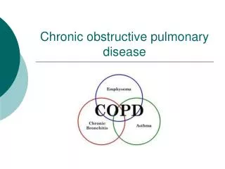
Definition Diseasecharacterized by persistentairflowlimitationthatis usuallyprogressive and associatedwithanenhancedchronic inflammatoryresponse in theairways and thelung to inhaled noxiousparticles or gases. Chronic expiratory flow limitation - maximum expiratory flows - slow forced emptying of the lungs Combination of two pathological processes - airways damage - emphysema
Chronic bronchitis defined in clinical terms chronic cough with sputum production - (3 months a year, 2 successive years) - excluded cardiac or other pulmonary causes Emphysema definedanatomically permanent, destructiveenlagrement of airspacesdistal to theterminalbronchioleswithoutobviousfibrosis COPD
Chronic airflow limitation - mechanisms Chronicinflammationcauses: - structuralchanges and narrowing of thesmallairways (obstructivebronchiolitis) - destruction of thelungparenchymaleads to theloss of alveolarattachments to thesmallairways and decreasinglungelasticrecoil - thesechangesdiminishtheability of theairways to remainopenduringexpiration
Risk factors Cigarette smoking 1 - antitrypsindeficiency Solid fuel used for indoor heating or cooking without adequate ventilation Heavilypollutedenvironments
100 Never smoked 75 Smoked regularly Stopped at age 45 yrs FEV1 % 50 Disability 25 Stopped at age 65 yrs Death 0 75 25 50 Age yrs
Cellular and biochemical mechanisms Inflammation: alveolar macrophages, CD8+ T lymphocytes, neutrophils production of elastase, cathepsine G, collagenase oxidative stress in smokers and in COPD patients Neutrophil and macrophage enzymes and oxidants destroy components of extracellular matrix (collagen, elastin, fibronectine, proteoglycans) Loss of cellular components of lung parenchyma: - elastase can induce apoptosis - cells exposed to oxidants may undergo necrosis
Imbalance proteases antiproteases system oxidants antioxidants Small airways disorder Destruction of lung parenchyma
Pathology of peripheral airways • mucus plugging • goblet cell hyper/metaplasia • fibrosis • smooth muscle hypertrophy
Peak exp. flow Maximal expiratory efforts 12 6 V´ ( l.s-1 ) 0 - 6 25 50 75 100 0 VC ( % exhaled)
COPD phenotypes symptoms obstruction severity risk of exacerbations occurrence of comorbidities
Frequentcomorbidities Loss of weight Dysfunction of skeletal muscles Osteoporosis Progression of aterosclerosis and coronary artery disease