Download
1 / 23
250 likes | 1.61k Vues
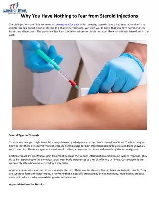
Steroid Joint injections . Updated 2010. Why inject joints?. Can be joint or soft tissue i.e. articular or periarticular Low risk e.g. septic arthritis occurs 1 in 40,000 Provide good symptom relief. Basic principles before you start. History and examination

E N D
Steroid Joint injections Updated 2010
Why inject joints? Can be joint or soft tissue i.e. articular or periarticular Low risk e.g. septic arthritis occurs 1 in 40,000 Provide good symptom relief
Basic principles before you start History and examination Try conservative treatment first e.g. physio, NSAIDs, orthotics and continue after joint injection. Careful patient selection Consent & provide ARCUK PILeaflet Know your anatomy! Undertake as few injections as possible to settle the problem, max 3-4 monthly (no more than 3 for tennis elbow per lifetime)
Indications for injection Osteoarthritis Rheumatoid arthritis Gouty arthritis Synovitis Bursitis Tendonitis Muscle trigger points Carpal tunnel syndrome
Inject with caution Reducing the risk of infection Never inject an infected joint. Avoiding injecting through infected skin or psoriatic plaques. Avoid injecting adjacent to infected skin/skin ulcers. Avoid injecting patient on concurrent oral steroids. Mediswabs or iodine should be used with a no touch or aseptic technique. Reducing the risk of bleeding If injecting weight bearing joints advise rest for 24 hours post injection. Don’t inject patients on warfarin Reducing the risk of tendon rupture Don’t inject near the Achilles tendon. Don’t inject into tendons.
Contraindication to injection Adjacent osteomyelitis or skin infection Bacteraemia Hemarthrosis Impending (scheduled within 3 months) joint replacement surgery Septic arthritis Joint prosthesis Osteochondral fracture Periarticular cellulitis / severe dermatitis/ soft tissue infection Plaque psoriasis at the injection point Poorly controlled diabetes mellitus Uncontrolled bleeding disorder or coagulopathy
Technique Complete the consent form and provide a Patient Information Leaflet prior to the procedure Inject the corticosteroid with as little pain and as few complications as possible. Do not attempt any injections in the vicinity of known nerve or arterial landmarks e.g. lateral epicondyle of elbow ok, medial – beware ulnar nerve Never inject into the substance of a tendon Sterile technique
Technique 2 ANTICIPATION! Get your kit ready ie: Needles, syringes, sterile container, LA, steroid, gloves, drapes, chlorhexidine, cotton wool, plaster. 1 or 2 needle technique (green to draw up and blue to give) Clean area – ensure solution has DRIED (esp iodine) prior to injecting
Technique 3 Always withdraw syringe back first to ensure not injecting into blood vessel Decide if you want to use lidocaine with the depomedrone Use a different needle to draw up (green) to the one you use to inject (blue or orange).
What doses of depo-medrone should you use? • Troc Bursitis 40-80mg • Knee 40-80mg • Shoulder 40mg • Tennis elbow 10-20 mg 9using a ‘peppering’ technique • +/- Lidocaine when injecting the shoulder or knee
What to warn the patient Pain returns after 2 hours, when the local anaesthetic wears off – may be worse than before. If pain is severe or increasing after 48hrs, seek advice Warn of local side effects Advise to seek help if systemic s/es develop
Local side effects Infection, subcutaneous atrophy, skin depigmentation, and tendon rupture (<1%). Post-injection ‘flare’ in 2-5% Often are the result of poor technique, too large a dose, too frequent a dose, or failure to mix and dissolve the medications properly. NB corticosteroid short duration of action – can be as short as 2-3 weeks relief.
Knee injections Patient on the couch, knee slightly bent Palpate superior-lateral aspect of patella Mark 1 fingerbreadth above + lateral to this site Clean LA, corticosteroid Clean + bandage
Plantar fasciitis Procedure painful + no evidence for long-term benefit Pt indicate tender spot Approach from thinner skin + direct posterior-laterally Small blelbs as near to bony insertion as possible Do not inject fascia itself
Shoulder injection • Glenohumeral joint • AC joint • Subacromial space • Long Head of Biceps • Older patients: 2-3 x/ year • Younger – consider surgery if no improvement (risk rotator cuff rupture)
Glenohumeral joint injection Pt sits, arm by side, externally rotated Find sulcus between head of humerus and acromion Posterolateral corner of acromion (2-3 cm inferior) Direct needle anteriorly toward coracoid process Insert needle to full length Fluid should flow easily
AC joint injection Palpate clavicle to distal aspect Slight depression where clavicle meets acromion Insert needle from anterior and superior approach Direct needle inferiorly
Sub-acromial joint injection Posterior and lateral aspect of shoulder Inferior to lower edge of posterolateral acromion Insert inferior to acromion at lateral shoulder Direct needle toward opposite nipple Insert needle to full length Fluid should flow easily
Elbow epicondyle injection Very effective in short term – 92% Benefits do not normally persist beyond 6 weeks Lateral (tennis elbow) + medial (golfer’s elbow) epicondylitis Patient supine
Tennis elbow (lateral) Arm adducted at side Elbow flexed to 45 degrees Wrist pronated Insert needle perpendicular to skin at point of maximal tenderness Insert to bone, then withdraw 1-2 mm Inject corticosteroid solution slowly
Golfer’s elbow (medial) Beware ulnar nerve! Rest arm in comfortable abducted position Elbow flexed to 45 degrees Wrist supinated Point of maximal tenderness - insert to bone, then withdraw 1-2 mm Inject corticosteroid solution slowly
De Quervain’s tenosynovitis Inflammation of thumb extensor tendons -Extensor pollicis brevis -Abductor pollicis longus Occurs where tendons cross radial styloid
De Quervain’s tenosynovitis Maximally abduct thumb (accentuates abductor tendon) Injection site Snuffbox at base of thumb Aim 30-45 degrees proximally toward radial styloid Insert needle between the 2 tendons (not in tendon) Do not inject if paraesthesias (sensory branch radial nerve)