Download
1 / 35
450 likes | 1.89k Vues
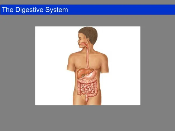
소화기계통 Digestive System. Oral cavity – pharynx – esophagus – stomach – small intestine – large intestine – rectum – anal canal – anus. Liver, pancreas. Oral Cavity( 입안 ). Vestibule( 입안뜰 ) Oral cavity proper( 고유입안 ). Palate ( 입천장 ) Hard palate( 단단입천장 ) Soft Palate( 물렁입천장 ). Tongue ( 혀 )

E N D
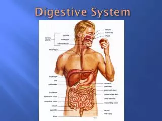
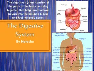
Oral cavity – pharynx – esophagus – stomach – small intestine – large intestine – rectum – anal canal – anus Liver, pancreas
Oral Cavity(입안) Vestibule(입안뜰) Oral cavity proper(고유입안)
Palate(입천장)Hard palate(단단입천장)Soft Palate(물렁입천장) Tongue(혀) Apex (혀끝) Root (혀뿌리) Dorsum (혓등) Margin (혀모서리) Inferior surface(혀아래면) Frenulum(혀주름띠) Papillae(유두) 성곽유두(vallate papilla), 버섯유두(fungiform papilla) 실유두(filiform papilla), 잎새유두(foliate papilla)
Tooth(치아) Deciduous Teeth(탈락치아, 젖니) ABDCE Permanent Teeth(영구치아, 간니) (6,1), 2,(4,3,5,7),8 anatomical crown 해부치아머리 clinical crown 임상치아머리 neck 치아목 root 치아뿌리 enamel 사기질 dentin 상아질 cementum 시멘트질 gingiva 잇몸 periodontal memb 치아주위조직 pulp cavity 치수공간 apical foramen 치아뿌리끝구멍
Pharynx(인두) Nasopharynx(코인두) auditory tube, pharyngeal tonsil, tubal tonsil Oropharynx(입인두) palatine tonsil Laryngopharynx(후두인두) piriform recess
Esophagus 인두끝 - 들문구멍 목부분 가슴부분 배부분 length 25cm, diameter2cm 위치 :목에서는 정중면의 왼쪽에 위치 대동맥활 부위에서는 정중면에 위치 대동맥활 아래에서는 다시 왼쪽에 위치 가슴대동맥은 식도의 왼쪽에 위치 여덟째 등뼈 높이에서 식도는 대동맥의 위로 지남
식도가 좁아진 부분 : 반지연골부위, 대동맥활 부위, 가로막을 뚫는 부위 15 22 27 40
Esophagus muscular layer of esophagus superior1/3; striated muscle middle1/3; mixed inferior1/3; smooth muscle
Stomach 음식물을 잘게 부수어 반액체 상태로 만듦. carida, fundus, body, pylorus cardial notch (들문패임); 식도와 위바닥 사이 pylorus; pyloric antrum,pyloric canal lesser & greater curvature, angular incisure(모패임) ► gastric fold; rugae (위주름) -수축했을 때 위점막에 생기는 긴 주름 -점막층, 근육층(세층), 장막층 ► bed of stomach (위자리) - 누워있을 때 위가 놓이는 부위 - 가로막의 왼지붕, 지라, 왼콩팥과 부신, 지라동맥, 이자, 가로잘록창자간막, 가로잘록창자
Duodenum 1st part of small intestine pylorus~ duodenojejunal flexure(2nd LV) 지름이 가장 넓고 고정됨 이자머리를 둘러싸는 C자형 ampulla(샘창자팽대); 날문에서 이어지는 창자간막에 붙어 있어 움직이는 위부분
Duodenum Superior part(첫째부분 5cm)- Descending part(둘째부분 7~10cm)- Transverse part(셋째부분;아래부분 6~8cm)- Ascending part(넷째부분 5cm)-
Duodenum Superior part (첫째부분 5cm)- 간과 쓸개주머니에 의해 덮여 있음 뒤쪽에 portal vein, common bile duct, superior mesenteric a., IVC 가 있음 Descending part (둘째부분 7~10cm)- 온쓸개관과 이자관(합쳐져 쓸개이자관팽대)이 뒤안쪽벽의 큰샘창자유두의 꼭지로 열림
Duodenum Transverse part (셋째부분;아래부분 6~8cm)- 아래대정맥과 대동맥, 셋째허리뼈의 앞을 수평으로 가로지름 위창자간막동맥,정맥, 빈창자와 돌창자의 창자간막이 수평부분의 앞을 지남 Ascending part (넷째부분 5cm)- 앞으로 휘어져 빈창자와 연결됨 샘창자걸이근에 의해서 지지됨 (suspensory m. of duodenum; Treitz lig.)
Jejunum and Ileum duodenojejunal flexure~ileocecal junction 약 6~7m, jejunum2/5, ileum3/5 jejunum은 주로 LUQ에 위치 ileum은 주로 RLQ에 위치
빈창자와 돌창자의 차이 JejunumIleum 돌림주름이 발달 돌림주름 미약 무리림프소절 x 무리림프소절 있다. 활꼴동맥이 뚜렷 x 활꼴동맥이 뚜렷 곧은동맥이 길다 곧은동맥이 짧다. 벽이 더 얇다. 창문(fenestra) 창자간막지방이 더 있다.
Large intestine cecum, appendix, ascending colon, transverse colon, descending colon, sigmoid colon, rectum, (anal canal)으로 구성 작은창자와의 차이점 1) tenia coli (잘록창자띠); 두꺼워진 세로근육 2) haustra (잘록창자팽대); 띠가 짧아서 3) omental appendices (복막주렁) 4) 속공간의 지름이 매우 크다.
Cecum and Appendix 막창자와 막창자꼬리 돌창자와의 연결부분 ileal orifice 돌막창자구멍 ileocolic lip 돌잘록창자입술 (위입술) ileocecal lip 돌막창자입술 (아래입술) frenulum 돌창자구멍주름띠; 가로주름 Appendix 6~10cm 막창자의 뒤안쪽벽에 붙어 있음 mesoappendix 막창자 뒤쪽에 놓이는 경우가 많다
Colon 잘록창자 ascending colon right colic flexure오른잘록창자굽이 hepatic flexure간굽이 transverse colon left colic flexure 왼잘록창자굽이 splenic flexure 지라굽이 descending colon sigmoid colon
Spleen 지라 biggestlymph oragan 복막에 싸여 LUQ에 위치 hilum; 혈관이 드나드는 곳 9th~11th rib 앞 left colic flexure의 위앞쪽 길이 12cm, 너비 7cm
Pancreas 이자 샘창자와 지라사이, 위의 뒤에 위치 endocrine gland, exocrine gland head, neck, body, tail uncinate process; 배쪽이자싹의 잔재
Pancreas Pancreatic duct 이자관 쓸개관과 합쳐져 팽대된 쓸개이자관팽대를 형성하여 샘창자 내림부위의 큰샘창자유두로 열림. 이자관조임근, 온쓸개관조임근, 쓸개이자관팽대조임근 Accessory pancreatic duct 덧이자관 작은샘창자유두로 열림 ►이자관과 덧이자관은 서로 연결
Liver 간 biggestorgan male1400g~1800g, female1200g~1450g 대부분 RUQ(right hypochondrium), 일부 LUQ 7th ~ 10th rib 글리코겐저장, 쓸개즙생성, 지방대사, 해독
Surface of liver-diaphragmatic surface 표면이 매끈하고 볼록한 지붕모양 round lig.(간원인대) falciform lig.(낫인대) coronary lig.(관상인대) triangular lig.(세모인대) bara area(무장막구역)
Surface of liver-visceral surface hilum; caudate lobe~quadrate lobe portal v. & hepatic a. 들어가고 hepatic duct가 나옴 (portal triad, 간세동이). 그외 간의 신경얼기나 림프관이 드나든다. *간샘창자인대 impressions gastric, duodenal, colic, renal impressions
Hepatic segmentation 간구역 간동맥과, 간문맥에서 독립된 혈액공급을 받고 정맥의 유출과 쓸개즙의 분비도 독립적으로 이루어짐. 낫인대, 간원인대틈새, 정맥관인대틈새에 의해 오른엽과 왼엽으로 나누어짐. 임상적으로는 아래대정맥오목과 쓸개오목을 잇는 가상선이 오른엽과 왼엽을 나누는 기준이 됨.
Biliarysystem bile juice; 지방의 소화를 도움. ► extrahepatic biliary system Rt., Lt., hepatic duct common hepatic duct cystic duct common bile duct Gall bladder 쓸개 7~10cm, 30~60ml base, body, neck 쓸개목은 쓸개주머니관으로 이어짐
Rectum(곧창자) Rectosigmoid junction(곧창자구불창자경계) Levator ani(항문올림근) Pectinate line(빗살선) Transverse rectal fold(가로곧창자주름))
Anal canal(항문관) Circular muscle layer Longitudinal muscle layer Levator ani muscle (항문올림근) Sphincterani internus (속항문조임근) Sphincterani externus (바깥항문조임근) Anal column(항문기둥) Anal sinus(항문굴) Anal valve(항문판막) Anal pecten(항문가리비)