Download

1 / 34
340 likes | 344 Vues
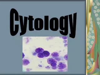
CYTOLOGY UNIT updated 2017. 2 NIPPLE DISORDERS & DISCHARGE. BY Soheir Mahfouz. Malignant smears. M. NEOPLASMS IN NIPPLE DISCHARGE & NIPPLE SCRAPES. Duct carcinoma : DCIS, medullary, papillary, comedo & colloid Paget ’ s disease Squamous cell carcinoma

E N D
CYTOLOGY UNIT updated 2017 Soheir Mahfouz-Self Study Series
2 NIPPLE DISORDERS & DISCHARGE BY Soheir Mahfouz Soheir Mahfouz-Self Study Series
Malignant smears Soheir Mahfouz-Self Study Series
M. NEOPLASMS IN NIPPLE DISCHARGE & NIPPLE SCRAPES • Duct carcinoma : DCIS, medullary, papillary, comedo & colloid • Paget’s disease • Squamous cell carcinoma NB: around 3% of malignant breast T. are associated with ND. A palpable mass /abnormal mammogram is usually found in such cases Soheir Mahfouz-Self Study Series
Cancer patients generally present with : A)Spontaneous rather than provoked discharge B)Unilateral & from 1 duct C)Grossly bloody D)May be associated with a breast mass or occurs in female over 40 years Soheir Mahfouz-Self Study Series
MICROSCOPIC FEATURES • Increased cellularity, • Tumour diathesis & a single cell population • Loose clusters / papillae with cells dropping out of the cluster giving an irregular border instead of a well defined one (loss of cohesion) Soheir Mahfouz-Self Study Series
MICROSCOPIC FEATURES • Malig. cytomorphology: • Cell & nuclear moulding • Large prominent nucleoli • Apple bite appearance due to irregularity in nuclear membrane • Hyperchromasia or excess palour & jagged chromatin Soheir Mahfouz-Self Study Series
NB: • When size of nuclei are double the size of an RBC consider SUSPICIOUS • Increased cellularity and older age group patient consider suspicious • Lymphocytes when present must be commented upon Soheir Mahfouz-Self Study Series
GROSS • Serous –Sanguinous - Serosanguinous Soheir Mahfouz-Self Study Series
WELL DIFFERENTIATED POORLY DIFFERENTIATED Cellularity ++ +++/ scanty Pattern Small clusters with loss of cohesion Papillae - Acini Small & large clusters with loss of cohesion Cells Single, small - medium cell population difficult to diagnose. Cells are minimally pleomorphic. Prominent nucleoli are not a feature Large cell population with ill defined boundaries. Cells are markedly pleomorphic Prominent nucleoli are a feature MICROSCOPIC Soheir Mahfouz-Self Study Series
Papilloma Young patient Carcinoma Old patient Background Clean+ RBC Dirty (tumour diathesis)+/- calcifications Pattern Hypocellular Clusters mostly tight Hypercellular Many discohesiveclusters DIFFERENTIAL DIAGNOSIS Soheir Mahfouz-Self Study Series
DIFFERENTIAL DIAGNOSIS BENIGN Vs MALIGNANT Soheir Mahfouz-Self Study Series
DIAGNOSIS & MANAGEMENT Soheir Mahfouz-Self Study Series
DIFFERENTIAL DIAGNOSIS Acute Mastitis Fibrocystic disease Early fat necrosis Inflammatory Inflammatory& lipopteinacious background Inflammatory Fat droplets PNLs Histiocytes Duct cells FC +/- PNLs Histiocytes Duct cells Foam cellsmany Apocrine cells PNLs Histiocytes Giant cells some lymphocytes( increase as the disease progresses ) & fat globules Soheir Mahfouz-Self Study Series
Benign Malignant Hormonal Mastitis Fibrocystic Papilloma Foam & duct cells +/- giant cells PNLs, pus cells, lympho histiocytes PNLs-Histiocytes Many foam cells Apocrine met Many duct cells Cohesive clusters Many duct cells Discohesive Tumour diathesis Milky/serous Pus/sanguinous Serous Serosanguinous Green cheesy Serous Serosanguinous Sanguinous Serous Serosanguinous Sanguinous DIFFERENTIAL DIAGNOSIS Soheir Mahfouz-Self Study Series
Benign Malignant Background Clean or dirty if inflammatory with sepsis Dirty (tumour diathesis) +/- calcification Pattern Polymorphic population Hypocellular few clusters mostly tight Monomorphic Hypercellular discohesive clustersMany Gross All types Serous-serosanguinous-sanguinous Cells No pleomorphism No cell or nuclear moulding No nucleoli or small Fine chromatin Many cell types Pleomorphism Cell & nuclear moulding Nucleoli big Coarse/ variable chromatin 1 cell type Soheir Mahfouz-Self Study Series
INTERPRETATION COMMENT & DIAGNOSIS 1) Unsatisfactory:Insufficient / degenerated epithelial cells RECOMMENDATIONS: repeat 2) Benign A) Inflammatory: Acute – subacute- chronic mastitis Soheir Mahfouz-Self Study Series
B) Epithelial proliferative lesion with NO ATYPIA • Hypercellular smears with no malignant features • Many ductal epithelial cells • Papillary structures(papilloma/papillomatosis) RECOMMENDATIONS: Excision biopsy • Fibrocystic disease can be diagnosed if a combination of the following cells are found: foam cells-apocrine cells- duct cells-RBCs-Inflammatory cells RECOMMENDATIONS: Follow up Soheir Mahfouz-Self Study Series
C)Atypical proliferative epithelial cell lesion Hypercellular smears with 1 / 2 features of neoplasia Lesions in this Mild category: Intraduct papilloma – atypical duct hyperplasia Marked: atypical duct hyperplasia & Insitu duct carcinoma =SUSPICIOUS RECOMMENDATIONS: Excision Biopsy Soheir Mahfouz-Self Study Series
Presence of atypical hyperplasia increases the RR of cancer development RR=4.9 This increases even more RR=18.1 with +ve family history Wrensh et al.1992,Am.J.Epidimiol.135:130-141 Soheir Mahfouz-Self Study Series
3)Suspicious/marked atypia: not all 4 criteria of malignancy are present This is generally reported when dealing with well differentiated tumours , marked atypical duct hyperplasia & Insitu duct carcinoma RECOMMENDATIONS: Excision biopsy 4) Malignant:Only when there is no doubt and all 4 features of malignancy are present: 1- Hypercellularity 2-discohesive clusters 3– neoplastic nuclear features 4- one cell population & a dirty smear(necrosis /calcification) RECOMMENDATIONS: Excision biopsy Surgery Soheir Mahfouz-Self Study Series
Ancilliary diagnostic methods Molecular Markers • Immunohistochemistry • Proteomics profiles by mass spectroscopy (protein finger printing) • Chromosomal alterations by FISH & other methods • RNA expression techniques (PCR) SEDE trial serial evaluation of ductal epithelium is a marker trial started 2004 ends 2007 Soheir Mahfouz-Self Study Series
MANAGEMENT OF BREAST DISCHARGE Soheir Mahfouz-Self Study Series
Patient management(in high risk group) Ductal lavage • Benign: repeat after 1-3 years • Mildly atypical: repeat after 1 year or consider prevention therapy • Markedly atypical/malignant: do ancillary diagnosis eg MRI, ductoscopy,or ductogram finally may require biopsy if a lesion is found DL results are not the basis for major surgical intervention more of a screening method in high risk groups O’Shaughnessy etal 2003,Surg.Clin.N.Am;83:753-769 Soheir Mahfouz-Self Study Series
MEDICAL TREATMENT & / follow up Inflammatory Hormonal Cysts SURGICAL EXCISION All proliferative samples (with or without atypia) Malignant smears Management Soheir Mahfouz-Self Study Series
THINGS TO REMEMBER (A) • ND is commonly benign in origin • Pregnancy & a variety of medical conditions & medications can cause milky discharge • Bilateral ND is almost always due to endocrinal problems • Discharge characters associated with high risk of underlying malignancy are: Unilateral-One duct-breast mass & bloody Soheir Mahfouz-Self Study Series
THINGS TO REMEMBER (B) • Bilateral multi duct secretion –ve Guiac is not malignant regardless of colour (milky-brown-green-blue-clear) • Cytology should not be performed on nipple discharge that is not grossly bloody or guiac +ve • A sticky straw coloured transparent discharge is characteristic of intraductal papilloma(Guiac +ve) • Cytology is useful in differentiating proliferative from inflammatory lesions in patients with Occult blood in discharge. Proliferative=surgery Inflammatory = medical Soheir Mahfouz-Self Study Series
FNAC Shirley2004,Uptodate12(2)(800)998-6374.(781)237-4788 Soheir Mahfouz-Self Study Series
Soheir Mahfouz Fahima Habib Zeinab Moheidin Essam Ayad Hossam Ahmed Sammar El Sheikh Ahmed Soliman Shady Anis Nada Iskandar Amira El Zahid Amany Mamdouh Abla Sayed Sherine Farouk Shaimaa Bebars CYTOLOGY UNIT members responsible for material collection
Material obtained from • Cytology unit, Kasr Al Ainy image bank • Cytosleuth http://www.bccancer.bc.ca/HPI/CE/cytotechnology/cytosleuthquiz/default.htm • 2000 The Johns Hopkins University School of Medicinehttp://www.hopkinsbreastcenter.org/pathology/malignant http://pathology2.jhu.edu/cyto_tutorial/Index.cfm • http://endeavor.med.nyu.edu/path-cases/breast/home/breastfnahome.html • http://dpalm.med.uth.tmc.edu/cytopath/cytologyimages.htm • Gail et al. 1989 • National Surgical Adjuvant Breast and Bowel Project (NSABP) revised this model in 1992 (model 2) • Vogel, 2004 diag. Cytopathol : 30: 1151-157 • Wrensh et al.1992,Am.J.Epidimiol.135:130-141 • www.miraeobgy.com/clinic03/breastcancer/ • www.breastcancerupdate.com/breast_cancer...files/ductal_lavage.html • emedicine.medscape.com/article/347305-overview • http://www.cancernews.com/images/aimages/kim/figure2.jpg • http://www.ncc.go.jp/sap-cc/NetWork/DAILY3/DC2001-108/DC2001-108-2.jpg • http://www.ncc.go.jp/sap-cc/NetWork/DAILY3/DC2001-108/DC2001-108-6.jpg • http://www.ncc.go.jp/sap-cc/NetWork/DAILY3/DC2001-108/DC2001-108-8.jpg • http://www.sap-cc.org/NetWork/DAILY3/DC2001-019/DC2001-19-1.JPG • Kini SR: color atlas of differential diagnosis in exfoliative and aspiration cytopathology 2nd ed p726 Lippincot Williams and Wilkens 2011 Self study series Breast Soheir Mahfouz
http://www.sap-cc.org/NetWork/DAILY3/DC2001-019/DC2001-19-2.JPGhttp://www.sap-cc.org/NetWork/DAILY3/DC2001-019/DC2001-19-2.JPG • UpToDate, Rose, BD (Ed), UpToDate, Waltham, MA, 2006. • Bernstein JL et al Cancer Res 2005 Sep 15;11(18):6528-35 • Harris JR, et al., eds. Diseases of the breast. 2d ed. Philadelphia: Lippincott Williams & Wilkins, 2000:38. • Krishnamurthy et.al., 2002, Diagnostic Cytopath . 27(5)::261-264 • H. Zakhour and C. Wells, Diagnostic Cytopathology of the Breast, Churchill Livingstone, London, UK, 1999. • Wrensh et al.1992,Am.J.Epidimiol.135:130-141 • www.healthline.com • http://www.sap-cc.org/NetWork/DAILY5/DC2003-456/DC2003-456-04.jpg • http://www.sap-cc.org/NetWork/DAILY5/DC2003-456/DC2003-456-07.jpg • http://www.sap-cc.org/NetWork/DAILY5/DC2003-456/DC2003-456-02.jpg • http://rds.yahoo.com/S=96062883/K=nIPPLE+DISCHARGE/v=2/SID=e/l=IVS/SIG=12duoas1t/EXP=1106820648/*-http%3A//www.stonybrookhospital.com/BALDWIN/image/papil.jpg • http://www.sap-cc.org/NetWork/DAILY/DC2000-18/DC2000-18-5.jpg • http://www.sap-cc.org/NetWork/DAILY/DC2000-18/DC2000-18-7.jpg • http://www.sap-cc.org/NetWork/DAILY/DC2000-18/DC2000-18-3.jpg • http://pathresidents.com/usapathology/em-handbook/introduction-section/utility-cases/uc-figs-fnab1.html • http://www.ncc.go.jp/sap-cc/NetWork/DAILY3/DC2001-058/DC2001-058-07.JPG • http://www.ncc.go.jp/sap-cc/NetWork/DAILY2/DC2000-195/DC2000-195-01.JPG • http://www.breastcancerupdate.com/breast_cancer_symposium/poster_graphics/MARKED ATYPICAL SUSPICIOUS.jpg • Http://www.sap-cc.org/NetWork/DAILY2/DC2000-205/DC2000-205-02.JPG • http://www.sap-cc.org/NetWork/DAILY2/DC2000-205/DC2000-205-01.JPG • http://www.sap-cc.org/NetWork/DAILY2/DC2000-205/DC2000-205-06.JPG • http://www.sap-cc.org/NetWork/DAILY2/DC2000-205/DC2000-205-04.JPG • O’Shaughnessy etal 2003,Surg.Clin.N.Am;83:753-769 • http://www.cancernews.com/images/aimages/kim/figure2.jpg • http://www.sap-cc.org/NetWork/DAILY/DC2000-18/DC2000-18-3.jpg Soheir Mahfouz-Self Study Series