Download
1 / 49
510 likes | 758 Vues
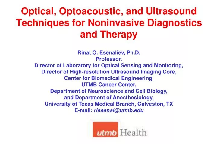
Optical, Optoacoustic, and Ultrasound Techniques for Noninvasive Diagnostics and Therapy. Rinat O. Esenaliev, Ph.D. Professor, Director of Laboratory for Optical Sensing and Monitoring, Director of High-resolution Ultrasound Imaging Core, Center for Biomedical Engineering,

E N D
Optical, Optoacoustic, and Ultrasound Techniques for Noninvasive Diagnostics and Therapy Rinat O. Esenaliev, Ph.D. Professor, Director of Laboratory for Optical Sensing and Monitoring, Director of High-resolution Ultrasound Imaging Core, Center for Biomedical Engineering, UTMB Cancer Center, Department of Neuroscience and Cell Biology, and Department of Anesthesiology, University of Texas Medical Branch, Galveston, TX E-mail: riesenal@utmb.edu
Our Group Has Pioneered Noninvasive Therapeutic and Diagnostic Technologies: • Optoacoustic Platform for Noninvasive Sensing, Monitoring and Imaging: absorption contrast • Noninvasive Monitoring with Optical Coherence Tomography (OCT): scattering contrast • Nanoparticles and Radiation (Optical, Ultrasound) for Cancer Therapy or for Drug Delivery: Including laser + gold nanoparticle (nanoshells, nanorods, etc.) for cancer therapy
Publications, Patents, Grants • 40 peer-reviewed papers • 15 patents including 7 issued patents • $9.3M in 23 research grants from NIH, DOD, state and private funding agencies
Research Team Collaborators: Donald S. Prough, M.D., Department of Anesthesiology, UTMB Michael Kinsky, M.D., Department of Anesthesiology, UTMB Claudia Robertson, M.D, Baylor College of Medicine, Houston Luciano Ponce, M.D., Baylor College of Medicine, Houston Joan Richardson, M.D., Department of Pediatrics, UTMB B. Mark Evers, M.D., Department of Surgery, UTMB Donald E. Deyo, D.V.M., Department of Anesthesiology, UTMB Douglas S. Dewitt, Ph.D., Department of Anesthesiology, UTMB Visiting Scientists: Valeriy G. Andreev, Ph.D., Physics Department, Moscow State University Alexander I. Kholodnykh, Ph.D., Physics Department Moscow State University Research Associates, Post-Doctoral Fellows, and Graduate and Undergraduate Students: Christian Bartels, M.S.; Saskia Beetz, B.S.; Peter Brecht, Ph.D.; Olga Chumakova, Ph.D., Inga Cicenaite, M.D.; Olaf Hartrumpf, B.S.; Dominique Hilbert, B.S.; Manfred Klasing, B.S.; Anton Liopo, Ph.D., Roman Kuranov, Ph.D.; Kirill V. Larin, Ph.D.; Irina V. Larina, Ph.D.; Margaret A. Parsley, B.S.; Igor Patrikeev, Ph.D.; Andrey Y. Petrov, B.S.; Yuriy E. Petrov, Ph.D.; Irina Y. Petrova, Ph.D.; Emanuel Sarchen, B.S.; Veronika Sapozhnikova, Ph.D.; Alexandra A. Vassilieva, M.D.; Karon E. Wynne, B.S.
Optoacoustic Grants NIH R01 # EB00763 “Novel Sensor for Blood Oxygenation”. NIH R21 # NS40531 “Optoacoustic Monitoring of Cerebral Blood Oxygenation”. NIH R01 # NS044345 “Optoacoustic Monitoring of Cerebral Blood Oxygenation”. John Sealy Memorial Endowment Fund for Biomedical Research. Grant: “Noninvasive Monitoring with Novel, High-resolution Optical Techniques”. Moody Center for Traumatic Brain & Spinal Cord Injury Research. Seed Grant: “Noninvasive Optoacoustic Hemoglobin Monitor” – Subaward from Noninvasix, Inc. Texas Emerging Technology Fund (TETF): “Noninvasive Platform for Blood Diagnostics” – Subaward from Noninvasix, Inc. NIH STTR: “Noninvasive Optoacoustic Monitoring of Circulatory Shock”. DOD: "Noninvasive Monitoring of Cerebral Venous Saturation in Patients with Traumatic Brain Injury“. DOD: “Noninvasive Circulatory Shock Monitoring with Optoacoustic Technique”.
Noninvasix, Inc. • UTMB Incubator Startup • Exclusive, world-wide license on optoacoustic monitoring, sensing, and imaging in humans and animals in vivo and in vitro (non-cancerous appl.) • Licensed key US and International patents including patents on monitoring OxyHb, THb, ICG, etc. in blood vessels and in tissues • FD: UTMB and Drs. Esenaliev and Prough are co-owners of Noninvasix
Optoacoustics for Biomedical Imaging, Monitoring, and Sensing - 1 Early 1990s: First Peer-reviewed Papers on Biomedical Optoacoustics Institute of Spectroscopy, Russian Academy of Sciences: R.O. Esenaliev, A.A.Oraevsky, V.S.Letokhov and A.A. Karabutov (Moscow State University)
Optoacoustics for Biomedical Imaging, Monitoring, and Sensing - 2 Since Mid 1990s we continued the biomedical optoacoustic works in the USA: Mid 1990s: Optoacoustic Signals from Deep Tissues (Depth: 5 cm) Late 1990s: First Optoacoustic Images Mid 1990s - Present: Optoacoustic Imaging, Monitoring and Sensing Patents: imaging, monitoring of temperature, coagulation, freezing, oxygenation, hemoglobin, other important physiologic parameters, etc.
Optoacoustics for Biomedical Imaging, Monitoring, and Sensing - 3 2001: We Obtained First High-resolution Optoacoustic Images Photonics West/ BIOS/SPIE Statistics: At present, Biomedical Optoacoustics is the fastest growing and largest area in biomedical optics
MOTIVATION - 1Cerebral Venous Oxygenation Monitoring: for Patients with Traumatic Brain Injury (TBI) and Cardiac Surgery Patients • Traumatic brain and spinal cord injuries are the leading cause of death • and disability for individuals under 50 years of age (car accidents, falls, etc.) • 150,000 patients/year with moderate or severe traumatic brain injury • and 2 million/year with total TBI (mild, moderate, severe). • Clinical data indicate that low cerebral venous blood oxygenation (below 50%) results in worse outcome (death or severe disability); 55-75% is normal (venous!) • Continuous and accuratemonitoring of cerebral venous blood oxygenation is critically important for successful treatment of these groups of patients • Existing methods are invasive (catheters in jugular bulb), • and noninvasive (NIRS) cannot measure cerebral venous oxygenation
MOTIVATION - 2Central Venous Oxygenation Monitoring: for Patients with Circulatory Shock • Circulatory shock is common in critically ill patients • Clinical data indicate that low central venous blood oxygenation (below 70%) results in worse outcome (death or severe complications); 70% is normal • Continuous and accuratemonitoring of central venous blood oxygenation is critically important for successful treatment of these patients: reduction in mortality from 46.5% to 30% • Existing methods are invasive (pulmonary artery catheters), while noninvasive (NIRS) cannot measure central venous oxygenation
MOTIVATION - 3THb Monitoring Total hemoglobin concentration ([THb]) measurement/monitoring is important clinical test during: Routine health assessment (reveals anemia - [THb] < 11 g/dL or polycythemia [THb] > 18 g/dL ) 2 billion people suffer from anemia worldwide Surgical procedures involving rapid blood loss, fluid infusion, or blood transfusion Existing methods are invasive: Blood sampling Optical monitoring in an extracorporeal blood circuit Noninvasive methods are inaccurate: Pressing need for noninvasive methods for continuous, accurate [THb] measurement
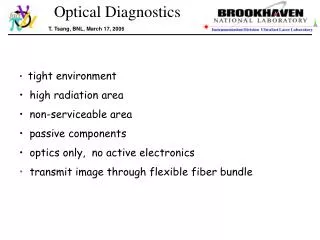
Pure Optical Techniques (NIRS) Cannot Detect Signals Directly from Blood Vessels Due to Strong Light Scattering in Tissues
Optoacoustic Technology:Optical Contrast + Ultrasound Resolution • Light pulses into blood in a vessel • Blood hemoglobin absorbs light & emits ultrasound in proportion to concentration in the vessel (due to thermal expansion) • Ultrasound wave travels without scattering and arrives at specific time proportional to blood vessel depth • Sensor detects ultrasound • Software determines location, size, and oxygenation of blood in the vessel Optical Input Acoustic Output
Thermoelastic (Optoacoustic) Pressure, P: DT– laser-induced temperature rise; µa – absorption coefficient; F– fluence of the laser pulse; r –density; cv – heat capacity at constant volume Principle of Laser Optoacoustic Monitoring and Imaging Laser Optoacoustic Monitoring and Imaging Is Based on Generation, Detection, and Analysis of Thermoelastic Pressure Waves Induced by Short Laser Pulses
High Resolution and Contrast Can Be Achieved Only when Short Laser Pulses Are Used (The Condition of Stress Confinement) L – desirable spatial resolution tac– time of propagation of acoustic wave through the distance = L tac = L/ cs where cs = 1.5 mm/ns – speed of sound in tissue The Condition of Stress Confinement: where tlaser– laser pulse duration
Generation of Optoacoustic Wave in Absorbing Medium Spatial Distributionof Optoacoustic Pressure in an Absorbing Medium without Scattering: • [1/oC] – thermal expansion coefficient; cs [cm/s] – speed of sound; Cp [J/goC] – heat capacity at constant pressure; F(z) [J/cm2] – fluence of the optical pulse; µa [cm-1] – absorption coefficient of the medium; G –Grüneisen parameter (dimensionless) Since: Temporal Profile of Optoacoustic Waves in the Medium:
Generation of Optoacoustic Wave in Tissue Spatial Distributionof Optoacoustic Pressure in a Tissue (not close to the surface): k–coefficient depending on tissue optical properties µeff – tissue attenuation coefficient Temporal Profile of Optoacoustic Waves in the Tissue:
Advantages of Optoacoustic Technique • High Contrast (as in Optical Tomography) because • It Utilizes Optical Contrast • High Resolution (as in Ultrasonography) due to • Ultrasound Wave Detection • (Insignificant Scattering of Ultrasonic Waves • Compared with Light Wave Scattering in Tissues)
Absorption Spectra of Oxy- and Deoxyhemoglobin Steven L. Jacques, Scott A. PrahlOregon Graduate Institute
Therapeutic Window: 600 – 1400 nm Low absorption and low scattering = Deep penetration Steven L. Jacques, Scott A. PrahlOregon Graduate Institute
Our Goal Is to Develop Optoacoustic Devicefor Monitoring: • Oxygenation • Cerebral • Central Venous • Peripheral Venous • Arterial • Total Hb Concentration • Pathologic Hemoglobins • Carboxyhemoglobin • Methemoglobin • Dye Concentration (ICG) • Blood Volume • Cardiac Output • Hepatic Function • Noninvasive Venous Pressure • Noninvasive Arterial Pressure
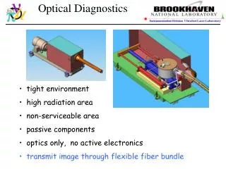
Optoacoustic Monitoring Systems Used in these Studies:OPO-Based Optoacoustic Monitoring Systems andLaser Diode-Based, Optoacoustic Monitoring Systems Wavelengths: 680 – 2400 nm; Duration: 10 - 150 ns Optoacoustic Probes Used in these Studies:Single-element Probes,Focused Probes,Optoacoustic Arrays Specially developed sensitive, wide-band ultrasound detectors: 25kHz – 10 MHz
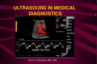
Ultrasound Imaging Systems Used in these Studies:Standard Clinical GE SystemsandSiteRite SystemsHigh-Resolution Vevo System
High Resolution: 30 microns at depth of up to 25 mm Real-time Longitudinal Studies Measure Physiological parameters Contrast/Molecular Imaging Translatable to man Novel, High-resolution Ultrasound Imaging System (Vevo, VisualSonics)
Outcome after Head Injury Closely Correlates with Cerebral Venous Oxygenation / OxyHb Saturation Below 50%: death or severe disability Schell RM et al., Anesth Analg 2000;90:559. Gopinath SP, Robertson CS, Contant CF, et al. Jugular venous desaturation and outcome after head injury. J. Neurol. Neurosurg. Psychiatry. 57:717-23, 1994.
Noninvasive, optoacoustic monitoring of cerebral venous blood oxygenation
Noninvasive, Optoacoustic Cerebral Venous Blood Oxygenation Monitoring in Sheep 1064 nm 700 nm
Optoacoustic Spectra from Human SSS and Hemoglobin Absorption Spectra
Ultrasound Imaging and Corresponding Optoacoustic Signals from Central Veins
Optoacoustic Spectra from Carotid Artery and Central Vein Carotid Artery Central Vein
Continuous, Real-time Measurement of Central Venous Oxygenation Using Optoacoustic Monitoring System (Stable Subject) <SO2> = 75.1 +/- 1.1 %
High-Resolution Ultrasound Imaging and Corresponding Optoacoustic Signals from Peripheral Veins
Optoacoustic Spectra from Peripheral Vein and Radial Artery and Hemoglobin Absorption Spectra Radial Artery Peripheral Vein
Optoacoustic Signals from Bloodin Radial Artery Phantom Optoacoustic signal from sheep blood at different concentrations of THb (gradual dilution of blood) and its amplitude
Conclusions • The optoacoustics is a platform monitoring and imaging technology with high (optical) contrast and high (ultrasound) resolution in tissues • The sensitive, wide-band optoacoustic probes provide sufficient lateral and axial resolution for measurements in large and small blood vessels • The optoacoustic monitoring systems may provide clinically acceptable accuracy of cerebral, central, and peripheral venous oxygenation measurements • The optoacoustic monitoring systems may provide high accuracy of hemoglobin measurements
Nanoparticles and Radiationfor Cancer Therapy or for Drug Delivery
Overview of Technology Chemotherapy, surgery, radiation therapy have limitations and are not capable of safe and efficient therapy of solid tumors. This technology offers efficient cancer therapy with no or minimal side effects. The technology is based on interaction of light, microwaves, radiowaves, or ultrasound with nanoparticles. Radiation + Nanoparticles Nanoparticle-mediated Therapy Drug Delivery Light-Induced Therapy MW-Induced Therapy RW-Induced Therapy US-Induced Therapy Light-Enhanced DD MW-Enhanced DD RW-Enhanced DD US-Enhanced DD
Drug Delivery Problem Approximately 1.4 million new cases are diagnosed and more than 500,000 deaths occur as a result of cancer every year in the United States. Many promising therapeutic agents have been proposed for cancer therapy for the past two decades. Their potential is proven in numerous preclinical studies. However, limited success has been achieved in tumor therapy. Barriers to drug delivery: • blood vessel wall • interstitial space • cancer cell membrane Penetration is especially poor for macromolecular therapeutic agents: • monoclonal antibodies 150 – 300 kDa • cytokines 6 – 70 kDa • antisense oligonucleotides 5 – 10 kDa • gene-targeting vectors > 1,000 kDa Modified from R.Jain, “Barriers to drug delivery in solid tumors”, Scientific American, 1994.
INTERACTION of NANOPARTICLES with ULTRASOUND CAN ALTER the BARRIERS to DRUG DELIVERY • The nanoparticles can be selectively accumulated in tumors by: • • “passive” delivery due to increased leakage of tumor capillaries (EPR effect) • • “active” delivery with the use of antibodies, short peptides, etc. • Interaction of particles with radiation produce cavitation and other effects • Which RESULTS IN: • • rupture or changes in tumor blood vessel wall and cancer cell membrane • • microconvection in the interstitium
Advantages of PLGA nanoparticles: can accumulate in tumors (EPR effect) biodegradability, biocompatibility, may provide stable cavitation, PLGA approved for clinical use by FDA (surgical sutures, etc.) Biodegradable and biocompatible polymer Poly (D,L-lactide-co-glycolic acid) 50:50, PLGA The nanoparticles was prepared by double water/oil/water emulsion solvent evaporation technique followed by filtration with 220-nm Millex filters
PLGA Nanoparticles Optical Microscopy SEM High Performance Particle Sizer HPPS 5001 Zetasizer Nano
PLGA nanoparticles (0.5% solution) Definity (0.2% solution)
In Vivo Gene Delivery Control Tumor Irradiated Tumor
Precise Damage Induced by Ultrasound+Nanoparticles Deeply in Tissues
(whole tumor) (Necrotic region) Reference subtracted Contrast Images Reference subtracted Wash in curve whole tumor area Reference subtracted Wash in curve Necrotic region
Wash in curve for nanoparticles circulating in tumor Reference subtracted Contrast Image
Acknowledgement • John Sealy Memorial Endowment Fund • 2. Texas Advanced Technology Program • (grant #004952-0088-2001) • 3. Department of Defense Breast Cancer Research Program • (grant #DAMD17-01-1-0416) • 4. National Institutes of Health • (grant #RO1 CA104748) • 5. Department of Defense Prostate Cancer Research Program • (grant #W81XWH-04-1-0247)