Download

1 / 66
900 likes | 1.56k Vues
Antigen – Antibody Reactions or Serological Reactions. Antigen-Antibody interactions. Characterized as : Non-covalent interaction (similar to “lock and key” fit of enzyme-substrate) Do not lead to irreversible alteration of Ag or Ab

E N D
Antigen-Antibody interactions Characterized as: • Non-covalent interaction (similar to “lock and key” fit of enzyme-substrate) • Do not lead to irreversible alteration of Ag or Ab • This exact and specific interaction has led to many immunological assays that are used to: • detect Ag or Ab • diagnose disease • measure magnitude of humoral IR • identify molecules of biological and medical interest
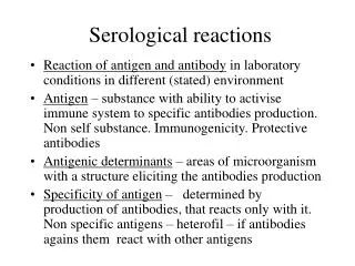
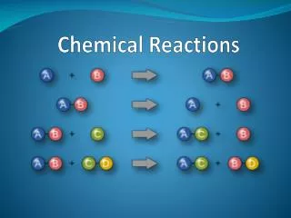
Introduction • Ag – Ab reactions are one of the most specific noncovalent biochemical reactions known • The forces that hold the reactants together are: - van der Waals forces - Electrostatic forces - Hydrophobic forces • They can be represented by the simple formula: Ag + Ab ↔ AgAb • The reaction is driven to the right but it is reversible
Strength of Reaction • The strength of the reaction (how far it is driven to the right) is referred to as affinity • Antibody affinity -A quantitative measure of binding strength - Combined strength of the noncovalent interactions between a binding site on an Ab & monovalent Ag - Affinity varies broadly among immunoglobulins
Strength of Reaction • Antibody avidity -Avidity is often used to describe the collective affinity of multiple binding sites on an antibody molecule - True strength of the Ab -Ag interaction within biological systems - The interaction at one site will increase the possibility of reaction at a second site - High avidity can compensate for low affinity (secreted pentameric IgM has a higher avidity than IgG )
CROSS REACTIVITY • Antibody elicited by one Ag can cross-react with a related Ag. • Occurs if two different Ags share identical or very similar epitope 1- Vaccinia virus and smallpox virus 2- Rabies & JE vaccine 3- Streptococcus pyogenes infection: heart & Kidney damage following infection 4- Original antigenic sin. 5- Bacterial Ag and sugars on RBC
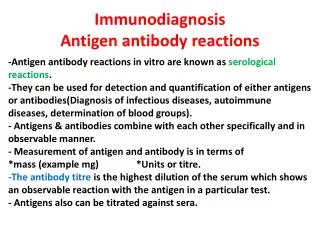
STAGES OF Ag - AbREACTIONS • Primary reactions Vs secondary reactions: Small Ag - Ab complexes Vs large complexes (The Lattice hypothesis) • Development of macroscopic manifestations reactions (e.g. immunoprecipitation) • Ag – Ab reactions involving IgM are confined to the blood stream, while those of lower molecular weight (IgG and IgE) can leave the vasculature and enter tissues • Time required is hours to days for precipitin formation leading to irreversible immunoprecipitates
LATTICE THEORY • Lattice formation (visible Ag - Ab aggregates) occurs when: • Ag is multivalent (contains more than 2 identical epitopes) • Cross-linking of Ags by specific Abs (2 or more antigen-binding sites) • Molar ratios of epitopes and antigen-binding sites are optimal (zone of equivalence)
Zone of equivalence
LATTICE THEORY • Zones of lattice formation • Far Ag excess (no ppt. formed; free Ag in supernatant) -- “postzone” • Ag excess (sub-optimal ppt.; free Ag in spnt.) • Zone of equivalence (maximum ppt.; no Ag or Ab in spnt.) • Ab excess (sub-optimal ppt; Ab in spnt.) • Far Ab excess (no ppt; Ab in spnt.) -- “prozone”
ZONES OF PRECIPITIN FORMATION Precipitin Curve
METHODS THAT DETECT Ag- Ab REACTIONS • Primary Reactions: - Immunofluorescence (IF) - Radioimmunoassay (RIA) - Enzyme immunoassay (EIA) - Immunonephelometry (measures picogram to nanogram quantities of analyte) • Secondary Reactions - Agglutination Techniques - Precipitation Techniques ± Electrophoresis
Precipitation • Precipitation can take place in capillary tubes, test tubes, and in gel
Precipitation in gel - Double diffusion - Single (radial) diffusion - Combination of diffusion in gel and electrophoresis
SINGLE VS. DOUBLE DIFFUSION • Single diffusion • Supporting medium (gel) contains one reactant at a uniform concentration • Only the unknowns move through the medium • Double diffusion • Gel is inert (contains no reactants) • Both Ag and Ab travel through the medium
The region of equivalence
RADIAL IMMUNODIFFUSION • Ab uniformly distributed in gel; Ag diffuses outward from a well (single diffusion) • Ag- Ab complexes form as concentric rings around the well at zone of equivalence • At a set time, ring diameters are measured • [Ag] is directly proportional to the ring d2 • Unknown value is determined by comparing to a 3-standard curve
RADIAL IMMUNODIFFUSION Samples Standards Precipitin Rings a b c ABC Standard Curve
RADIAL IMMUNODIFFUSION • Fahey method (kinetic) • Read at 18 hours • Plot [std] vs. ring diameter on semi-log paper • Mancini method (endpoint) • Read at 48 or 72 hours • Plot [std] vs. ring diameter squared on graph paper • Results reliable only if the ring size is within the range of the standards; if greater than highest std, dilute and repeat test • Used to measure IgM, IgG, C4,C3,transferrin, CRP, others
OUCHTERLONY DOUBLE DIFFUSION • Ag & Ab placed in wells cut into an agarose gel (both reactants diffuse) • Precipitin line (or arc) indicates Ab has specificity for Ag • Position of precipitin between wells depends on MW and concentration of reactants • 3 possible patterns of reaction: identity, non-identity, partial identity
OUCHTERLONY DOUBLE DIFFUSION Ouchterlony PlatesPrecipitin Patterns
ELECTROIMMUNOASSAY (ROCKET) • Electrophoresis hastens movement of Ag (placed in wells) through Ab -imbedded gel (single diffusion) • Selected pH (8.6) keeps Abs at their isoelectric point; they will not move • Rocket-shaped precipitin bands will form at zone of equivalence (changes as reactants move) • [Ag] proportional to length of rocket • Unknowns compared to standards
ELECTROIMMUNOASSAY (ROCKET) • May be used to quantitate plasma proteins such as coagulation factors, alpha-fetoprotein, C3, C4, CRP, haptoglobin • Compared with RID: • faster • similar sensitivity • requires electrophoretic equipment and more technological finesse • Largely replaced by immunonephelometry
IMMUNONEPHELOMETRY • Ag + Ab AgAb microscopic Ag - Ab complexes • Microcomplexes cause light moving through the suspending solution to scatter • Nephelometer detects light scattered at a 90o angle • Amount of light scattered at 90o is proportional to Ag - Ab complexes formed • Sensitive and quantitative technique used for measurement of many serum proteins
IMMUNOELECTROPHORESIS (IEP) • Electrophoresis and double diffusion • 2 stages • Proteins separated by electrophoresis • Antiserum placed in trough parallel to separated proteins; all reactants diffuse in all directions • Precipitin forms at zones of equivalence • Trough may be filled with simple or complex antisera yielding simple to complex patterns
33 Immunoelectrophoresis of normal human serum.
IMMUNOELECTROPHORESIS (IEP) • Qualitative to semi-quantitative • Serum, urine, or CSF may be analyzed • Complex patterns may be difficult to interpret • Useful to detect: • missing proteins • abnormal proteins • normal proteins in abnormal concentrations • Used to evaluate conditions such as multiple myeloma • Largely replaced by immunofixation
IMMUNOFIXATION ELECTROPHORESIS (IFE) • Proteins that were separated by electrophoresis are exposed to Ab directly, instead of through diffusion • Steps: • Electrophoresis of protein mixture in gel (use serum or urine samples) • Paper strips imbedded with specific Ab are “blotted” onto gel; Ags transfer to paper and bind to Abs • Strips washed (unbound material washes away) • Strips stained to reveal precipitin bands
IMMUNOFIXATION ELECTROPHORESIS (IFE) • Used to detect the presence of Igs in conditions like multiple myeloma • Fairly sensitive - Ab is highly specific, electrophoresis leaves Ag isolated and accessible • Faster and easier to interpret than IEP • Only 1 Ab may be used per strip
WESTERN BLOTTING • Similar to IFE but the unknown is Ab rather than Ag • Steps: • Separation of complex antigenic material (eg., viral proteins) by electrophoresis • Separated components transferred from gel to nitrocellulose paper by “blotting” • Unknown (or control) sera (which may have Abs) incubated with paper strips; Ag - Ab complexes ppt. at site of transfer • Strips washed; staining reveals complexes
FLOCCULATION Immunoprecipitation (or agglutination) of insoluble particles Characterized by very sharp pro- and postzones No precipitin formed in zones of Ab or Ag excess, only in zone of equivalence Clinically important examples, VDRL and RPR tests (screening tests for syphilis)
Flocculation Tests • VDRL (Venereal Disease Research Lab.) test • RPR (Rapid Plasma Reagin) test
Agglutination • Titer • Zeta potential • Types of Agglutination - Direct agglutination or hemagglutination - Indirect (passive) agglutination or hemagglutination - Agglutination or hemagglutination inhibition • The Coombs test - Direct - Indirect
Agglutination • Qualitative slide agglutination • - identification of bacteria with antisera directed against • O, H, K antigens
Agglutination • Latex agglutination • Coagglutination
Agglutination • Tube agglutination tests: • -Gruber-Widal: typhoid fever (S. typhi) • - Weil-Felix: typhus (Rickettsia) • - Wright: brucellosis • Identify and titrate antibodies in the patient’s • serum. • Titre: is defined as the reciprocal of the • highest dilution of serum showing agglutination.