Download
1 / 12
120 likes | 204 Vues
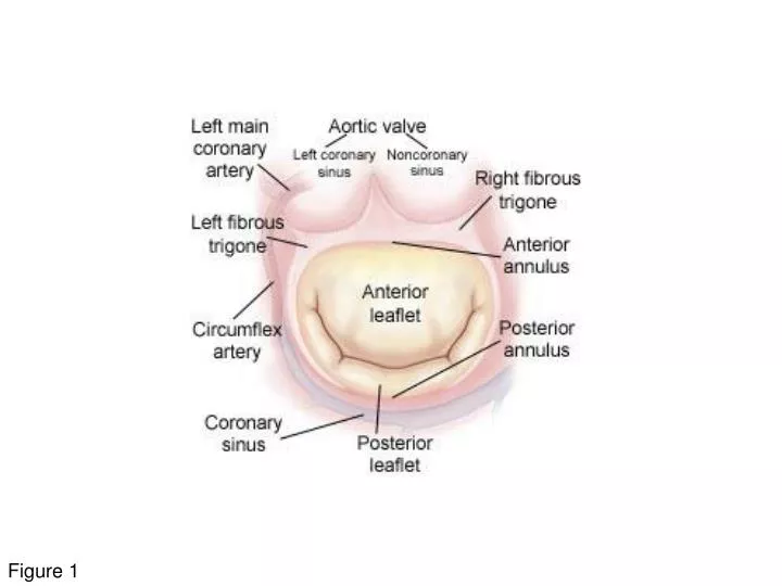
Figure 1. Figure 2. Figure 3. A1. P1. A2. A3. P2. P3. A. B. P3. A3. P2. A2. A1. P1. Figure 4. A1. A3. A2. P3. P1. P2. Figure 5. A2-P2. Ao. Med. A. A3. A1. A2. P3. P1. Lat. P2. P3-A2-P1. Ao. Med. B. A3. A1. A2. P3. P1. Lat. P2. P2-A2. Ao. Med. C.

E N D
A1 P1 A2 A3 P2 P3 A B P3 A3 P2 A2 A1 P1 Figure 4
A1 A3 A2 P3 P1 P2 Figure 5
A2-P2 Ao Med A A3 A1 A2 P3 P1 Lat P2 P3-A2-P1 Ao Med B A3 A1 A2 P3 P1 Lat P2 P2-A2 Ao Med C A3 A1 A2 P3 P1 Lat P2 P3-A3,A2,A1 Ao Med A3 A1 D A2 P3 P1 Lat P2 Figure 6
First Order Second Order Third Order Figure 10
Type I –normal leaflet motion • Annular dilatation • Leaflet perforation • Type II – increased leaflet motion • Ruptured chord • Ruptured papillary muscle • Elongated chord • Type III A – systolic and diastolic leaflet restriction • Commisural fusion • Leaflet thickening • Fused chordae • Type III B – systolic leaflet restriction • Leaflet tethering Figure 12