Download
1 / 47
520 likes | 1.01k Vues
Chapter 6 Bone and Skeletal Tissue. Part 2 Angela Peterson-Ford, PhD apetersonford@yahoo.com. 6. Bones and Skeletal Tissues Part 2. Stages of Intramembranous Ossification. An ossification center appears in the fibrous connective tissue membrane

E N D
Chapter 6Bone and Skeletal Tissue Part 2 Angela Peterson-Ford, PhD apetersonford@yahoo.com
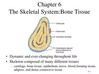
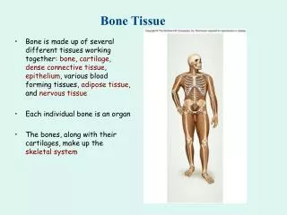
6 Bones and Skeletal Tissues Part 2
Stages of Intramembranous Ossification • An ossification center appears in the fibrous connective tissue membrane • Bone matrix is secreted within the fibrous membrane • Woven bone and periosteum form • Bone collar of compact bone forms, and red marrow appears
Stages of Intramembranous Ossification Figure 6.7.1
Stages of Intramembranous Ossification Figure 6.7.2
Stages of Intramembranous Ossification Figure 6.7.3
Stages of Intramembranous Ossification Figure 6.7.4
Endochondral Ossification • Begins in the second month of development • Uses hyaline cartilage “bones” as models for bone construction • Requires breakdown of hyaline cartilage prior to ossification
Stages of Endochondral Ossification • Formation of bone collar • Cavitation of the hyaline cartilage • Invasion of internal cavities by the periosteal bud, and spongy bone formation • Formation of the medullary cavity; appearance of secondary ossification centers in the epiphyses • Ossification of the epiphyses, with hyaline cartilage remaining only in the epiphyseal plates
Stages of Endochondral Ossification Secondary ossification center Articular cartilage Epiphyseal blood vessel Spongy bone Deteriorating cartilage matrix Hyaline cartilage Epiphyseal plate cartilage Spongy bone formation Primary ossification center Medullary cavity Bone collar Blood vessel of periosteal bud Formation of bone collar around hyaline cartilage model. 1 Cavitation of the hyaline cartilage within the cartilage model. 2 Invasion of internal cavities by the periosteal bud and spongy bone formation. 3 Formation of the medullary cavity as ossification continues; appearance of secondary ossification centers in the epiphyses in preparation for stage 5. 4 Ossification of the epiphyses; when completed, hyaline cartilage remains only in the epiphyseal plates and articular cartilages 5 Figure 6.8
Postnatal Bone Growth • Growth in length of long bones • Cartilage on the side of the epiphyseal plate closest to the epiphysis is relatively inactive • Cartilage abutting the shaft of the bone organizes into a pattern that allows fast, efficient growth • Cells of the epiphyseal plate proximal to the resting cartilage form three functionally different zones: growth, transformation, and osteogenic
Functional Zones in Long Bone Growth • Growth zone – cartilage cells undergo mitosis, pushing the epiphysis away from the diaphysis • Transformation zone – older cells enlarge, the matrix becomes calcified, cartilage cells die, and the matrix begins to deteriorate • Osteogenic zone – new bone formation occurs
Long Bone Growth and Remodeling • Growth in length – cartilage continually grows and is replaced by bone as shown • Remodeling – bone is resorbed and added by appositional growth as shown
Long Bone Growth and Remodeling Figure 6.10
Appositional Growth of Bone Central canal of osteon Periosteal ridge Penetrating canal Periosteum Artery Osteoblasts beneath the periosteum secrete bone matrix, forming ridges that follow the course of periosteal blood vessels. As the bony ridges enlarge and meet, the groove containing the blood vessel becomes a tunnel. 1 The periosteum lining the tunnel is transformed into an endosteum and the osteoblasts just deep to the tunnel endosteum secrete bone matrix, narrowing the canal. 2 As the osteoblasts beneath the endosteum form new lamellae, a new osteon is created. Meanwhile new circumferential lamellae are elaborated beneath the periosteum and the process is repeated, continuing to enlarge bone diameter. 3 4 Figure 6.11
Hormonal Regulation of Bone Growth During Youth • During infancy and childhood, epiphyseal plate activity is stimulated by growth hormone • During puberty, testosterone and estrogens: • Initially promote adolescent growth spurts • Cause masculinization and feminization of specific parts of the skeleton • Later induce epiphyseal plate closure, ending longitudinal bone growth
Bone Remodeling • Remodeling units – adjacent osteoblasts and osteoclasts deposit and resorb bone at periosteal and endosteal surfaces
Bone Deposition • Occurs where bone is injured or added strength is needed • Requires a diet rich in protein, vitamins C, D, and A, calcium, phosphorus, magnesium, and manganese • Alkaline phosphatase is essential for mineralization of bone • Sites of new matrix deposition are revealed by the: • Osteoid seam – unmineralized band of bone matrix • Calcification front – abrupt transition zone between the osteoid seam and the older mineralized bone
Bone Resorption • Accomplished by osteoclasts • Resorption bays – grooves formed by osteoclasts as they break down bone matrix • Resorption involves osteoclast secretion of: • Lysosomal enzymes that digest organic matrix • Acids that convert calcium salts into soluble forms • Dissolved matrix is transcytosed across the osteoclast’s cell where it is secreted into the interstitial fluid and then into the blood
Importance of Ionic Calcium in the Body • Calcium is necessary for: • Transmission of nerve impulses • Muscle contraction • Blood coagulation • Secretion by glands and nerve cells • Cell division
Control of Remodeling • Two control loops regulate bone remodeling • Hormonal mechanism maintains calcium homeostasis in the blood • Mechanical and gravitational forces acting on the skeleton
Hormonal Mechanism • Rising blood Ca2+ levels trigger the thyroid to release calcitonin • Calcitonin stimulates calcium salt deposit in bone • Falling blood Ca2+ levels signal the parathyroid glands to release PTH • PTH signals osteoclasts to degrade bone matrix and release Ca2+ into the blood
Hormonal Mechanism Figure 6.12
Response to Mechanical Stress • Wolff’s law – a bone grows or remodels in response to the forces or demands placed upon it • Observations supporting Wolff’s law include • Long bones are thickest midway along the shaft (where bending stress is greatest) • Curved bones are thickest where they are most likely to buckle
Response to Mechanical Stress • Trabeculae form along lines of stress • Large, bony projections occur where heavy, active muscles attach
Response to Mechanical Stress Figure 6.13
Bone Fractures (Breaks) • Bone fractures are classified by: • The position of the bone ends after fracture • The completeness of the break • The orientation of the bone to the long axis • Whether or not the bones ends penetrate the skin
Types of Bone Fractures • Nondisplaced – bone ends retain their normal position • Displaced – bone ends are out of normal alignment • Complete – bone is broken all the way through • Incomplete – bone is not broken all the way through • Linear – the fracture is parallel to the long axis of the bone
Types of Bone Fractures • Transverse – the fracture is perpendicular to the long axis of the bone • Compound (open) – bone ends penetrate the skin • Simple (closed) – bone ends do not penetrate the skin
Common Types of Fractures • Comminuted – bone fragments into three or more pieces; common in the elderly • Spiral – ragged break when bone is excessively twisted; common sports injury • Depressed – broken bone portion pressed inward; typical skull fracture • Compression – bone is crushed; common in porous bones
Common Types of Fractures • Epiphyseal – epiphysis separates from diaphysis along epiphyseal line; occurs where cartilage cells are dying • Greenstick – incomplete fracture where one side of the bone breaks and the other side bends; common in children
Common Types of Fractures Table 6.2.1
Common Types of Fractures Table 6.2.2
Common Types of Fractures Table 6.2.3
Stages in the Healing of a Bone Fracture • Hematoma formation • Torn blood vessels hemorrhage • A mass of clotted blood (hematoma) forms at the fracture site • Site becomes swollen, painful, and inflamed Hematoma Hematoma formation 1 Figure 6.14.1
Stages in the Healing of a Bone Fracture • Fibrocartilaginous callus forms • Granulation tissue (soft callus) forms a few days after the fracture • Capillaries grow into the tissue and phagocytic cells begin cleaning debris External callus New blood vessels Internal callus (fibrous tissue and cartilage) Spongy bone trabeculae Fibrocartilaginous callus formation 2 Figure 6.14.2
Stages in the Healing of a Bone Fracture • The fibrocartilaginous callus forms when: • Osteoblasts and fibroblasts migrate to the fracture and begin reconstructing the bone • Fibroblasts secrete collagen fibers that connect broken bone ends • Osteoblasts begin forming spongy bone • Osteoblasts furthest from capillaries secrete an externally bulging cartilaginous matrix that later calcifies
Stages in the Healing of a Bone Fracture • Bony callus formation • New bone trabeculae appear in the fibrocartilaginous callus • Fibrocartilaginous callus converts into a bony (hard) callus • Bone callus begins 3-4 weeks after injury, and continues until firm union is formed 2-3 months later Bony callus of spongy bone Bony callus formation 3 Figure 6.14.3
Stages in the Healing of a Bone Fracture • Bone remodeling • Excess material on the bone shaft exterior and in the medullary canal is removed • Compact bone is laid down to reconstruct shaft walls Healing fracture Bone remodeling 4 Figure 6.14.4
Homeostatic Imbalances • Osteomalacia • Bones are inadequately mineralized causing softened, weakened bones • Main symptom is pain when weight is put on the affected bone • Caused by insufficient calcium in the diet, or by vitamin D deficiency
Homeostatic Imbalances • Rickets • Bones of children are inadequately mineralized causing softened, weakened bones • Bowed legs and deformities of the pelvis, skull, and rib cage are common • Caused by insufficient calcium in the diet, or by vitamin D deficiency
Homeostatic Imbalances • Osteoporosis • Group of diseases in which bone reabsorption outpaces bone deposit • Spongy bone of the spine is most vulnerable • Occurs most often in postmenopausal women • Bones become so fragile that sneezing or stepping off a curb can cause fractures
Osteoporosis: Treatment • Calcium and vitamin D supplements • Increased weight-bearing exercise • Hormone (estrogen) replacement therapy (HRT) slows bone loss • Natural progesterone cream prompts new bone growth • Statins increase bone mineral density
Paget’s Disease • Characterized by excessive bone formation and breakdown • Pagetic bone with an excessively high ratio of woven to compact bone is formed • Pagetic bone, along with reduced mineralization, causes spotty weakening of bone • Osteoclast activity wanes, but osteoblast activity continues to work
Paget’s Disease • Usually localized in the spine, pelvis, femur, and skull • Unknown cause (possibly viral) • Treatment includes the drugs Didronate and Fosamax
Developmental Aspects of Bones • Mesoderm gives rise to embryonic mesenchymal cells, which produce membranes and cartilages that form the embryonic skeleton • The embryonic skeleton ossifies in a predictable timetable that allows fetal age to be easily determined from sonograms • At birth, most long bones are well ossified (except for their epiphyses)
Developmental Aspects of Bones • By age 25, nearly all bones are completely ossified • In old age, bone resorption predominates • A single gene that codes for vitamin D docking determines both the tendency to accumulate bone mass early in life, and the risk for osteoporosis later in life