Download
1 / 43
430 likes | 609 Vues
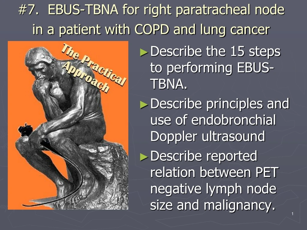
#7. EBUS-TBNA for right paratracheal node in a patient with COPD and lung cancer. Describe the 15 steps to performing EBUS-TBNA. Describe principles and use of endobronchial Doppler ultrasound Describe reported relation between PET negative lymph node size and malignancy.

E N D
#7. EBUS-TBNA for right paratracheal node in a patient with COPD and lung cancer • Describe the 15 steps to performing EBUS-TBNA. • Describe principles and use of endobronchial Doppler ultrasound • Describe reported relation between PET negative lymph node size and malignancy.
Case description(practical approach 7) • 67 year old male with a 50 pack- year history of smoking developed cough and weight loss (15kg) for six months. • Vital signs revealed a blood pressure of 160/80mmHg, heart rate 90/min, body temperature 37.2C and respiratory rate 18/min. • Physical examination shows prolonged expiratory breath sounds and egophony in right upper lung field. • He is a retired electrician and lives with his wife. He has no advance directives. • He desires all available active treatment modalities if diagnosed with cancer.
Case description(practical approach #7) • WBC 8000 (neutrophil 81%, lymphocyte 2%) • Hemoglobin 13 gm/dl, Platelets 310,000/mm3 • Arterial blood gas analysis pH 7.45, PaCO2 50 mmHg, PaO2 64 mmHg on 2L oxygen/min via nasal canula) • Pulmonary function tests revealed FEV1- 1.6 L (49% predicted), DLCO- 50% predicted
CT Chest: 3 cm right upper lobe mass. 1 cm right paratracheal lymph node is PET negative. CT guided transthoracic needle aspiration of the right upper lobe mass positive for non-small cell lung cancer. Case description(practical approach 7)
Initial Evaluation Procedural Strategies Techniques and Results Long term Management The Practical Approach • Examination and, functional status • Significant comorbidities • Support system • Patient preferences and expectations • Indications, contraindications, and results • Team experience • Risk-benefits analysis and therapeutic alternatives • Informed Consent • Anesthesia and peri-operative care • Techniques and instrumentation • Anatomic dangers and other risks • Results and procedure-related complications • Outcome assessment • Follow-up tests and procedures • Referrals • Quality improvement BI #. Practical Approach Title
Initial Evaluations • Exam • Prolonged expiratory phase • ECOG performance status 1 • Comorbidities • Severe COPD, HTN, Tobacco abuse • Support system • Wife and children all healthy and actively involved with patients care. • Patient preferences • Desires all available active treatment options.
Procedural Strategies • Indications: • Minimally invasive staging of non-small cell lung cancer with radiographically enlarged PET (-) node. • Contraindications: • None • Expected Results: sensitivity and NPV of EBUS 93.8% and 96.9% respectively* for NSCLC with lymph nodes of 5–20 mm on chest CT *Lee HS. Chest 2008; 134: 368–374.
Procedural Strategies • Risks-benefits: • EBUS-TBNA has no serious complications reported in the literature. • Agitation, cough, and presence of blood at puncture site have been reported infrequently.* • Same day procedure. • Cost savings when compared to mediastinoscopy.** • Increased risk in case general anesthesia required. *Eur Respir J 2009; 33: 1156–1164 **Gastrointestinal Endoscopy 69, No. 2, Supp 1, 2009, S260
Procedural Strategies • Therapeutic alternatives: • Endoscopic ultrasound difficult to access level 4 node compared with EBUS. In a head to head comparison* sensitivity and negative predictive value were 69% and 89% respectively) . • Mediastinoscopy gold standard. 78% sensitivity**, but requires general anesthesia. • VATS most invasive of alternatives. Only provides access to ipsilateral nodes. 75% sensitivity**. Benefits include definitive lobar resection at same time if frozen section negative. • Informed consent: • There were no barriers to learning identified. Patient has good insight into his disease and realistic expectations. *JAMA. 2008;299(5):540-546 **Chest 2007;132;202-220
Procedural Techniques and Results • Anesthesia and peri-operative care • Conscious sedation • Performed in clinic procedure room • Most commonly used drugs are midazolam and fentanyl • Cost savings when compared to OR and extra personnel required for general anesthesia • May make procedure more difficult for inexperienced operator • May be more appropriate for targeted biopsy than full staging of mediastinum* • Has been used in combined staging TBNA, EBUS, EUS procedures** *Chest 2008;134;1350-1351 **JAMA. 2008;299(5):540-546
Procedural Techniques and Results • Anesthesia and peri-operative care • General anesthesia with LMA • Mostly performed in OR but may be done in clinic • Total IV anesthesia with propofol is commonly used • LMA mask size 4 or 5 required • Allows easier biopsies of smaller nodes and complete mediastinal staging; better for less experienced operators • General anesthesia with ET tube • Size 8.5 in women and 9.0 in men • Allows for easier biopsies as above • Indications may include difficult LMA placement, obesity, and severe untreated GERD* • Causes EBUS scope to lie centrally in trachea • More difficult to visualize higher nodes *JCVA, Vol 21, No 6 , 2007: pp 892-896
Procedural Techniques and Results • Instrumentation • EBUS scope provides direct real time US imaging with curved array ultrasound transducer incorporated in distal end of bronchoscope • As of 09/2009, types of Scopes and US processors • Olympus- BF-UC160F-OL8 Hybrid scope • 2.0 mm working channel; 6.9 mm O.D • EU-C60 US processor 7.5 MHz with B-mode and color power doppler • Olympus BF-UC180F Hybrid scope • 2.2 mm working channel; 6.9 mm O.D. • ALOKA prosound US processor 5, 7.5, 10, 12 MHz and B, M, D-mode, flow and power flow modes • May also be used with EU-C60 processor • Pentax EB-1970UK Videoscope • 2.0 mm working channel; 6.3 mm O.D. • Hitachi HI Vision 5500 US processor 5MHz/7.5MHz/10MHz with B-mode and color Doppler
Aloka ProSound a5 Hitachi HI Vision 5500 EU-ME1
Procedural Techniques and Results • Instrumentation • Ultrasound processor • Adjustable gain and depth • Gain is the degree of brightness with which a given signal intensity is displayed. Analogous to a volume control knob on a stereo. • Depth- allows optimal display of an area of interest on the screen. • B mode and Doppler capabilities • B-mode (brightness mode) uses an array of transducers to scan a plane through the tissue to produce a two-dimensional image on the screen. • Doppler mode measures velocity of moving tissue. It detects blood flow in vessels and subsequently superimposes the display over the 2-D image.
Image quality adjustment GAIN CONTROL • Gain adjustments • The amplifier is often controlled by the operator of the instrument, who sets the gain for various depths of the tissue • Frequency adjustments • Higher frequency has better resolution but less depth of penetration Bronchoscopy International 15
Penetration Penetration: refers to the distance between an imaged area and the transducer. The time delay between the energy going into the body and returning to the US probe determines the depth from which the signal arises ( longer times= greater depths) Depth=velocity X time/2 Bronchoscopy International 16
Penetration and resolution Higher frequencies result in higher resolution. Higher frequencies (20 MHz) do not penetrate as deep as low frequencies (7.5 MHz). penetration frequency resolution Low frequencyhigh penetration Bronchoscopy International 17
Large transducers transmit powerful beams and increase penetration depth PLEURAL EFFUSION EBUS Penetration depth is less for EBUS than for thoracic ultrasound. Bronchoscopy International 18
Scanning methods Convex Transducer For the convex probe, the scanning plane is parallel to the scope Bronchoscopy International 19
BF-UC160F-OL8 Specifications http://www.olympusamerica.com/msg_section/download_brochures/b_bfuc160f_ol8.pdf Bronchoscopy International 20 Bowling MR, South Med J. 2008 May 101(5) 534-8
Procedural Techniques and Results • Instrumentation • Needle • Olympus NA-201SX-4022 or Medi-Globe SonoTip II • 22 gauge echogenic needle with stylet • Needle guide system locks to scope • Lockable needle and sheath • Precise needle projection up to 4 cm • Anatomic dangers and other risks • Major blood vessels- azygous, PA, aorta, SVC and Left atrium • Pneumothorax and pneumomediastinum • A case of bacterial pericardial effusion and nodal infection have recently been reported as complications following EBUS with full needle extension***. *Chest 2004;126;122-128 **Eur Respir J 2002; 19:356–373 ***Eur Respir J 2009; 33:935-938
Procedural Techniques and Results • Results and procedure-related complications • The 4R node was successfully sampled with EBUS under general anesthesia and a 9.0 cuffed endotracheal tube. • There was representative tissue on cytology and it was negative for malignancy. • There were no complications.
Long-term Management Plan • Outcome assessment • Patient underwent RUL lobectomy. Intraoperative mediastinal staging confirmed negative nodes. • At 1 month post operatively patient was back to preoperative baseline functional status. • Follow-up tests and procedures • Clinical evaluation every 3-6 months for the first 2 years with surveillance imaging every 6 months (CXR or CT)* • Referrals • He was also referred to oncology for consideration of adjuvant chemotherapy for I B disease. • Quality improvement • Early staging and definitive treatment of non-small cell lung ca • Expected 5 year survival for Stage Ib ~ 55%** *Chest 2007 132:355S-367S **Lung Cancer (2007) 55, 371-377
Procedure Technique Step 1 Advance needle through the working channel (neutral position) Step 2 Secure the needle housing by sliding the flange
Procedure Technique Step 3 Release the sheath screw Step 4 Advance and lock the sheath when it touches the wall
Procedure Technique Step 5 Release the needle screw Step 6 Advance the needle using the “jab” technique
Procedure Technique Step 7 Visualize needle entering target node Step 8 Move the stylet in and out a few times to dislodge bronchial wall debris.
Procedure Technique Step 9 Remove the stylet Step 10 Attach syringe
Procedure Technique Step 11 Apply suction Step 12 Pass the needle in and out of the node 15 times
Procedure Technique Step 13 Release suction by removing syringe Step 14 Retract the needle into the sheath
Procedure Technique • Step 15 Unlock and remove the needle and sheath and prepare smears.
Q 2: Describe principles and use of endobronchial Doppler ultrasound 33
Doppler ultrasound B-mode (brightness mode) uses an array of transducers to scan a plane through the tissue to produce a two-dimensional image on the screen. Doppler mode measures velocity of moving tissue. It detects blood flow in vessels and subsequently superimposes the display over the 2-D image. 34
Doppler ultrasound: Color Power Doppler Bronchoscopy International 35
Doppler Effect The frequency of the reflected ultrasound wave is changed when it strikes a moving object ( i.e blood in vessels)= Doppler effect Doppler frequency shift= ΔF= Ft-Fr=2 X Ft X (v/c) X cosθ Ft transmitted frequency, Fr received frequency, v speed of moving target, c speed of sound in soft tissue, θ angle between the direction of blood flow and direction of the transmitted sound phase Bronchoscopy International 36
4R Ascending aorta Bronchoscopy International 37
The Doppler angle needs to be 60 degrees or slightly less to the long axis of the vessel to obtain the correct velocity Strong Doppler signal is obtained when the scanning plane forms a sharp angle with the blood vessel • ΔF= Ft-Fr=2 X Ft X (v/c) X cos θ cosine(60 degrees) = 0.5 Bronchoscopy International 38
The Doppler angle needs to be 60 degrees or slightly less to the long axis of the vessel to obtain the correct velocity • ΔF= Ft-Fr=2 X Ft X (v/c) X cos θ Very weak or no Doppler signal is obtained when the scanning plane is perpendicular to the blood vessel cosine(90 degrees) = 0 Bronchoscopy International 39
Q 3: Describe reported relation between PET negative lymph node size and malignancy. 40
The size of PET (-) nodes impacts probability of malignancyMediastinal lymph nodes and relation with metastatic involvement: a Metanalysis. Langen et al, Eur J Cardiothorac Surg 2006;29:26-29 • Probability for malignancy in lymph nodes measuring 10-15 mm in the short axis is 29%,and about 60% if nodes are larger. • If nodes 10-15 mm and PET Negative, probability for malignancy is 5%. • Refrain from mediastinoscopy, proceed with thoracotomy • If nodes > 16 mm and PET Negative, probability for malignancy is 21%. • Proceed with mediastinoscopy
Bronchoscopy International: Practical Approach, an Electronic On-Line Multimedia Slide Presentation. http://www.Bronchoscopy.org/PracticalApproach/htm. Published 2007 (Please add “Date Accessed”). All efforts are made by Bronchoscopy International to maintain currency of online information. All published multimedia slide shows, streaming videos, and essays can be cited for reference as: Thank you
Prepared with the assistance of Steven Escobar MD and Septimiu Murgu MD www.bronchoscopy.org BI Practical Approach #1