Download

1 / 26
290 likes | 596 Vues
CNS DEVELOPMENT. Stages in Neural Tube Development. Neural plate. Neural folds. Neural tube. Time-Line. Formation of nervous system occurs during the embryonic stage: End of second week to end of eighth week. Time-Line. Superior (anterior or cranial) neuropore closes by day 27.

E N D
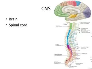
Stages in Neural Tube Development • Neural plate. • Neural folds. • Neural tube.
Time-Line • Formation of nervous system occurs during the embryonic stage: End of second week to end of eighth week.
Time-Line • Superior (anterior or cranial) neuropore closes by day 27. • Inferior (posterior or caudal) neuropore closes by day 30.
Subdivision of Cranial End of Neural Tube • Tripartite brain. • Pentapartite brain.
Tripartite Brain • Prosencephalon. • Mesencephalon. • Rhombencephalon.
Pentapartite Brain • Prosencephalon: Telencephalon (most anterior). Diencephalon. • Mesencephalon. • Rhombencephalon: Metencephalon. Myelencephalon.
Telencephalon Primordia • Lumina: Lateral ventricles (I, II). • Floor: Basal ganglia (nuclei). Olfactory lobes and nerves. • Roof: Cerebral hemispheres.
Diencephalon Primordia • Lumen: Third ventricle. • Roof: Epithalamus. • Walls: Thalamus. • Floor: Hypothalamus and infundibulum.
Mesencephalon Primordia • Lumen: Cerebral aqueduct (of Sylvius). • Roof =Tectum: Superior and inferior colliculi. • Floor: Tegmentum.
Metencephalon Primordia • Lumen: Part of fourth ventricle. • Roof: Cerebellum. • Floor: Pons.
Myelencephalon Primordia • Lumen: Rest of fourth ventricle. • Main part: Medulla oblongata. • Roof: Posterior choroid plexus.
Histogenesis of Neural Tube • Initial tube wall = Pseudostratified epithelium: Single layer of cells, but cells are of different heights. All cells are in contact with a basement membrane. • Outermost membrane = External limiting membrane.
Histogenesis of Neural Tube • Some neuroepithelial cells remain attached to the basement membrane and will form a single layer of ependymal cells that will line the entire ventricular system and the neural canal.
Histogenesis of Neural Tube • Tube differentiates into two concentric rings by day 26: Mantle layer and marginal layer.
Histogenesis of Neural Tube • Other cells lose contact with the basement membrane and will migrate past the ependymal cells to form a new outer layer of densely packed cells collectively called the: Mantle layer: • Cells that make up the mantle layer are: NEUROBLASTS. • Note that mantle layer is still covered by the external limiting membrane.
Histogenesis of Neural Tube • Neuroblasts in the mantle layer will begin to grow processes (axons) that will form a new outer layer: Marginal layer. The marginal layer is also located beneath the external limiting membrane. • The marginal layer will form the white matter of the spinal cord and the brain. • The mantle layer forms the gray matter of the brain and spinal cord (except for the cortices).
Anencephaly • Failure of cranial end of neural tube to close.
Arnold-Chiari deformity • Inferior cerebellum and medulla are elongated and protrude into vertebral canal. • Medulla and pons are small and deformed. • Hydrocephalus. • Malformation of lower cranial nerves: Deafness. Tongue, facial muscle, lateral eye movement weakness.
Spina Bifida Occulta • Results from a failure of the inferior neuropore to close. • Vertebral arch(-es) fails to develop in caudal area. • Spinal cord function is usually normal.
Spina Bifida Cystica • Characterized by a sac-like cyst at the caudal end of spine. • Spinal cord and/or meninges may be found in the cyst. • Spinal cord function may be impaired. • May be lower extremity dysfunction. • Bladder and bowel function may be impaired.
Meningocele • Form of spina bifida cystica. • Only meninges found in sac. • Spinal cord function may be impaired. • Signs and symptoms vary depending on location and severity of malformation.
Meningomyelocele • Form of spina bifida cystica. • Both meninges and spinal cord are found in sac. • Always results in abnormal growth of spinal cord. • Lower extremity paralysis. • Bowel and bladder dysfunction. • Loss of sensation to lower limbs.
Myeloschisis • Failure of caudal neural folds to close. • Most severe of the defects.
Holoprosencephaly • Failure of prosencephalon to divide into two cerebral hemispheres. • Often associated with facial deformities: Single orbit with two eyes or one eye or no eye. Proboscis-type nose located above eye. Cleft lip and palate.