Download
1 / 49
550 likes | 1.08k Vues
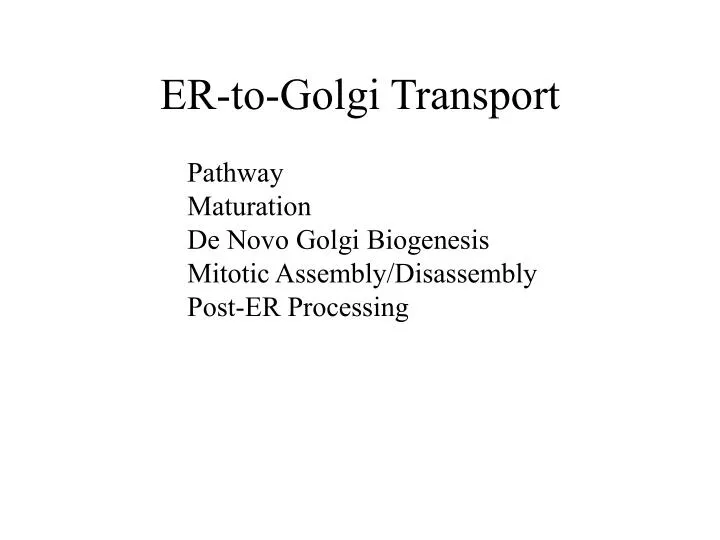
ER-to-Golgi Transport. Pathway Maturation De Novo Golgi Biogenesis Mitotic Assembly/Disassembly Post-ER Processing. Pathway Diagram. Sorting, Retention, Retrieval. ER. ERGIC. Golgi. The Golgi is the central processing and sorting station of the secretory pathway. ER. ERGIC. Golgi.

E N D
ER-to-Golgi Transport Pathway Maturation De Novo Golgi Biogenesis Mitotic Assembly/Disassembly Post-ER Processing
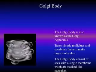
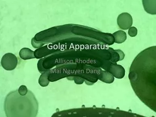
ER ERGIC Golgi The Golgi is the central processing and sorting station of the secretory pathway ER ERGIC Golgi
The Golgi maintains its complex structure in the presence of continuous membrane traffic trans medial cis
Enzymes in COPI? Table II. Relative lateral distribution of Golgi proteins and VSV-G within the Golgi complex Golgi stack (cisternae) Lateral rims (uncoated) Coated buds and vesicles Man II 68.9 ± 3.9 13.6 ± 2.7 17.5 ± 2.8 Giantin 58.2 ± 2.6 18.3 ± 2.8 23.5 ± 2.7 KDELr 35.8 ± 4.3 8.9 ± 2.0 55.3 ± 4.5 rBet1 25.6 ± 4.9 15.7 ± 4.0 58.7 ± 5.6 VSV-Gts045–GFP (20 min, 32°C) anti-GFP 93.1 ± 1.4 4.3 ± 1.3 2.6 ± 0.6 anti–VSV-G–lum 94.1 ± 0.8 3.4 ± 0.6 2.5 ± 0.5 ------------------------------------------------------------------------ Numbers represent the percentages (mean ± SEM) of the total labeling over the distinct membrane categories and were obtained by analyzing 50 Golgi complexes for each antibody.
COPI Vesicles Concentrate Enzymes not Cargo ------------------------------------------------------------------------ Markers Amount of vesicles (% of starting membranes) ------------------------------------------------------------------------ GTP GTPS arf-1 Q71L ------------------------------------------------------------------------ Golgi enzymes Mann II 13.2 ± 1.3 6.1 ± 1.4 4.2 ± 1.1 NAGT I 25.8 ± 2.7 3.1 ± 1.0 3.0 ± 0.8 GalT 11.0 ± 2.0 1.7 ± 0.4 1.8 ± 0.4 Anterograde cargo pIgR 4.6 ± 1.0 3.6 ± 1.4 3.3 ± 1.1 Phospholipids 2.7 ± 0.7 3.1 ± 0.7 2.5 ± 0.8 ------------------------------------------------------------------------
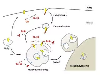
Trafficking may be sufficient for Golgi self-assembly Golgi ERGIC cis medial trans Trans Golgi Network Homotypic fusion MT COP I COP I COP I ER COP II
Can the Golgi self-assemble? Idea: Use a reversible ER export block -Brefeldin A to collapse Golgi -H89 to block ER export and collapse ERGIC
GM130 co-fractionates with ER membranes Golgi ER untreated BFA H89
min after H89 washout 0’ 4’ Re-emergence of the Golgi from the ER GM130 10’ 30’
Reassembled Golgi membranes are stacked BFA H89 30’ washout
Early compartmentalization of Golgi membranes after ER exit 5’ Giantin GPP130
Transport paths & consequences of their regulation Mitotic: vesicles=>block in docking Osmotic:ER=>ER export block
Blocking p115, but not giantin or GM130, causes vesiculation
What are the essential p115 interactions? (i.e. candidates for regulation at mitosis) acidic tail head domain coiled-coil domain Rab1 Giantin GM130 PLCg1 SNAREs phosphorylation site GBF1
100 80 60 40 mock 24 h 48 h 72 h 96 h 20 0 mock 96 h p115 siRNA induces time-dependent depletion of p115 GPP130 p115 p115 mock %intact p115 levels siRNA
siRNA-induced p115 depletion results in Golgi fragmentation p115 GPP130 mock siRNA
20 40 60 80 100% f +p115 Mismatched p115 rescues siRNA-induced Golgi fragmentation inj. marker p115 GalNac-T2 % injected cells with intact Golgi
O The giantin/GM130-binding domain is NOT required for Golgi biogenesis in vivo WT S941A Dtail Acidic Tail 20 40 60 80 100% Targeted? + WT + S941A + Dtail % expressing cells with intact Golgi
O WT Dcc1 The SNARE domain of p115 is required for Golgi biogenesis in vivo cc1 + + + + + ? + Targeted?
Working model for mechanism of action of p115 recruitment of p115 by Rab1 binding to t-SNARE catalysis of SNARE pairing p115 v-SNARE Rab1 t-SNARE => dissect SNARE mechanism w/ further mutagenesis => test other p115 interactions-> distinct phenotypes?
rbet1 syntaxin5 membrin sec22 Sub-regions of the cc1 domain show homology to distinct SNAREs rbet1 cc1 core complex domain syntaxin5 membrin p115 homology region sec22
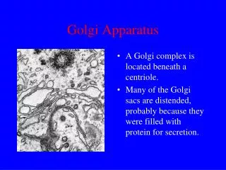
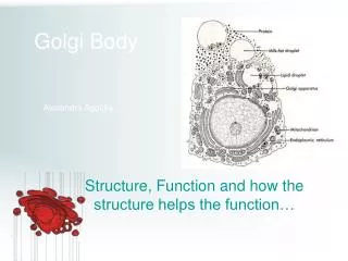
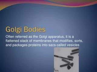
Part 1 Post-ER Processing Modification of carbohydrate side-chains Proteolytic activation of precursors
Processing Reactions endoH sensitive endoH resistant cis trans