Download
1 / 12
130 likes | 444 Vues
Viewing Microstructures of Materials with the Optical Microscope. Elizabeth Merten and Fumio Ohuchi University of Washington, Seattle Department of Materials Science & Engineering. Processing. Microstructure. Properties. Performance. Key Concepts.

E N D
Viewing Microstructures of Materials with the Optical Microscope Elizabeth Merten and Fumio Ohuchi University of Washington, Seattle Department of Materials Science & Engineering
Processing Microstructure Properties Performance Key Concepts Microstructures of a material relate to: • materials properties • mode of processing* • materials performance Microstructures for metallic samples can be resolved into individual crystal “grains” This phenomena can easily be observed in utilizing various characterization tools. One such tool is the Light Optical Microscopes (LOM) * variety of grain shape and size may be observed
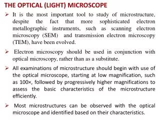
Optical Microscopy Optical microscopes are typically utilized for: • transparent samples (transmitted light microscopes) • metals such as brass, we must use a reflecting-type microscope* Optical microscopes allow us to magnify samples from 1-100X • Metal surfaces require special polishing and etching in order to resolve grains** • Photographs of microstructures are often called “micrographs” Micrographs of a Brass sample as well as a schematic of a basic transmitted light microscope are shown on the following slides *Reflected light microscope is similar but has the light source entering from above, through the viewing optics **Details for this process can be found in MatEd module “Brass Hardness”
Anatomy of an Optical Microscope • Eyepiece • Objective lens turret • Objective lens • Coarse adjustment knob • Fine adjustment knob • Stage for holding the sample • Light source reflected light microscope has light source entering from above • Light adjustment diaphragm, filters and condenser Figure 1: Basic Optical Microscope* *Image taken from Wikipedia :http://en.wikipedia.org/wiki/File:Optical_microscope_nikon_alphaphot_%2B.jpg
Micrographs of Brass Figure 1: Micrograph of brass as received, 50x Figure 2: Micrograph of brass 40% cold rolled, 50 x Can you see the elongation of the grains on the processed sample?
Magnification Image resolution Shortest distance between two points on a specimen that can still be distinguished by the observer Resolving power of microscopes are the most important feature • influences the ability of the system to distinguish between fine details Objective Lens—magnifies the object Eyepiece enlarges the image (does not add to resolution)
Resolution Available resolution depends on • Magnification of the objective lens • Uniform illumination of the sample • Numerical aperture (ability of the lens to collect light and resolve detail) • Wavelength of light * *shorter wavelengths are capable of resolving details to a greater degree than are the longer wavelengths
Numerical Aperture NA = n• sin(µ) Maximum angle is 90º, so max (sinµ) = 1 n = index of refraction n (air) = 1.0003 n (water) = 1.33 n (oils) = up to 1.515 Maximum magnification is about 1000 NA, or • 1000x in air • 1500x in oils Figure 2: Diagram of an angular aperture* *Image taken from: http://www.microscopy.fsu.edu/primer/anatomy/numaperture.html
Can we get higher Magnification? Utilizing higher magnification objectives or eye pieces often displays degraded images and poor resolution (similar to missing pixels when a photo is enlarged too much) Higher magnification also reduces the depth of focus (also called depth of field) • Higher depth of focus • Shorter wavelengths • Low Magnification • Poor depth of focus • High Magnification Figure 3: Depth of Focus for a Single Lens * *Image taken from: http://ion.asu.edu/descript_opt.htm
Wavelength Visible range of light is 390-760 nm • Resolving power of a light microscope is roughly 0.2 µmeters • Under typical conditions it is quite difficult to obtain an image down to this resolution Electron microscopes utilize electrons • Resolves images up to 10-10 meters! • Obtain images with increased magnification and depth of focus
Remember… The main concept is that the resolution of the image is directly related to the useful magnification of the microscope and the perception limit of specimen detail.
References Wikipedia: Optical Microscope image • http://en.wikipedia.org/wiki/File:Optical_microscope_nikon_alphaphot_%2B.jpg Numerical Aperture image • http://www.microscopy.fsu.edu/primer/anatomy/numaperture.html Depth of Focus image • http://ion.asu.edu/descript_opt.htm