1 / 10
100 likes | 103 Vues
https://neuros.creative-biolabs.com/cell-culture-models.htm

E N D
Histology & Immunohistochemistry Services https://neuros.creative-biolabs.com Tel: 1-631-357-2254 Mail: info@creative-biolabs.com
Creative Biolabs Every vision. One solution
Histology & Immunohistochemistry Services Your goal is discovery. Our goal is to get you there faster. Creative Biolabs’ histology and immunohistochemistry services are custom developed and fully optimized to meet your needs in neuroscience preclinical research. Creative Biolabs leads the industry in pathology expertise and innovative automated technology that provides you with the highest quality endpoints complimentary to in vivo and ex vivo study results. We complete the tissue collection from one of our highly characterized tumor bank models, tissue processing, paraffin embedding, cutting and staining all in-house. Our Creative Biolabs Quality Control process ensures unparalleled results on each slide, every time. PATHOLOGY EXPERTISE Industry Leaders in Neuroscience Preclinical Histology & Immunohistochemistry We provide the high-quality results you need through: ACCESS FFPE TISSUE TMAs from Over 800 Characterized Tumor Bank Models Automation - Our tech-enabled workflows provide accelerated and reproducible histopathology data. Digitization- You’ll receive crisp, high-resolution images that can be viewed online instantly and live indefinitely on the cloud. Expertise- All of our services are monitored and reviewed by experts with decades of histology and pathology experience. HIGH QUALITY RESULTS Excellence in Quality Control Leading to Distinct Results We can help you process: Take your research to the next level with our end-to-end histology platform. Our workflow maximizes your data organization and usage while providing a pathway for collaboration with other researchers around the world. Embedded tissue Unstained slides Stained slides Digital slides Wet tissue >
Creative Biolabs’ Histology & Immunohistochemistry Services Creative Biolabs has digitized more than 350,000 slides for 3,000 researchers across 500 organizations >
Tissue Processing and Slide Generation Tissue processing and slide generation are critical steps in your research, and they require significant time and expertise. Our streamlined, expert-led workflow provides a fast and reliable solution for accelerated results: Gross, process, embed • Automated histology with fast, reliable results Unstained slide generation • Automated staining • We provide various staining methods, including: H&E • Trichrome • Alcian Blue • Toluidine Blue • Picrosirius Red • Periodic Acid Schiff • >
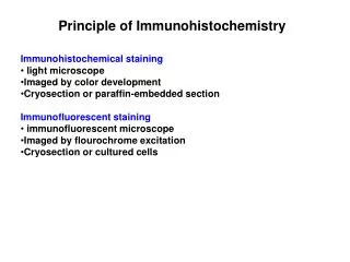
Immunostaining Our immunostaining service makes it fast and easy to visualize your proteins of interest. With an automated workflow that enhances reproducibility and speed of high-throughput studies, our visualization process saves you significant time in your research. We’re continuing to expand our extensive catalog of 350+ antibodies, validated on the Leica Bond system, for human, mouse, and various other species. If your marker has not yet been validated, our experts will optimize conditions for the best staining. Elevated immunostaining Immunohistochemical (IHC) and immunofluorescence (IF) staining allows for protein visualization. 400+ antibodies, validated on the Leica Bond system, for various tissues from human, mouse, and other species We can perform and customize IHC and IF staining for your research needs • • >
Slide Management Instantly view, manage, and share your data on the cloud. Creative Biolabs digitizes every stained slide with whole slide imaging (WSI) and stores them on PathologyMap, an intelligent, cloud-based platform that provides: Image analysis consultation Unlimited storage Online viewing Annotations Sharing Pathology network Organization Collaboration PathologyMap hosts the largest collection of preclinical data that fuels AI-driven QC tools. Equipping your workflow with our platform maximizes your data’s quality and usage. >
Pathologist Scoring Pathology expertise at your fingertips Access the world’s largest network of pathologists that provides objective assessment and on-demand diagnosis with the click of a button. We bridge the gap between preclinical researchers and pathologists with expertise from clinical human and veterinary studies. Pathologists review your whole slide images and provide assessments via an integrated report in industry-leading turnaround times. Meet our pathologists: 60 25,000 slides reviewed 80+ specialties represented board-certified pathologists averaging 16 years of experience 800+ 11 studies performed and reports generated toxicologic pathologists for GLP studies >
Image analysis Our service doesn’t end with beautiful images. Creative Biolabs equips researchers with a team of image analysis experts who provide objective assessment and measurable results. With HALO, Visiopharm, and AI tools, our team can help answer your research questions to accelerate your decisions and discoveries. Leverage your data with quantitative image analysis We provide quantitative solutions to inform your research questions. In addition to developing novel algorithms, our most established analyses include: • Number and percentage of positive cells • Differences within specific cellular subcompartments (such as nuclear_x0002_to cytoplasmic ratio) • Biologically relevant subregions within whole slide area (such as tumor from necrosis) • Differences in biomarker intensity and staining area >
https://neuros.creative-biolabs.com/ Mail: info@creative-biolabs.com Tel: 1-631-357-2254