Download
1 / 12
120 likes | 268 Vues
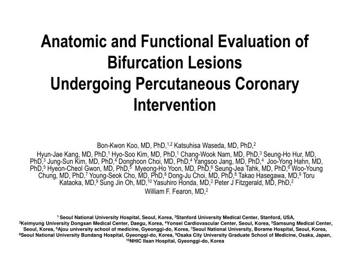
Anatomic and Functional Evaluation of Bifurcation Lesions Undergoing Percutaneous Coronary Intervention. Bon-Kwon Koo, MD, PhD, 1,2 Katsuhisa Waseda , MD, PhD, 2

E N D
Anatomic and Functional Evaluation of Bifurcation LesionsUndergoing Percutaneous Coronary Intervention Bon-Kwon Koo, MD, PhD,1,2KatsuhisaWaseda, MD, PhD,2 Hyun-Jae Kang, MD, PhD,1Hyo-Soo Kim, MD, PhD,1 Chang-Wook Nam, MD, PhD,3Seung-Ho Hur, MD, PhD,3 Jung-Sun Kim, MD, PhD,4DonghoonChoi, MD, PhD,4Yangsoo Jang, MD, PhD,4Joo-Yong Hahn, MD, PhD,5Hyeon-CheolGwon, MD, PhD,5Myeong-Ho Yoon, MD, PhD,6Seung-JeaTahk, MD, PhD,6 Woo-Young Chung, MD, PhD,7 Young-Seok Cho, MD, PhD,8 Dong-JuChoi, MD, PhD,8 Takao Hasegawa, MD,9 Toru Kataoka, MD,9 Sung Jin Oh, MD,10 Yasuhiro Honda, MD,2 Peter J Fitzgerald, MD, PhD,2 William F. Fearon, MD,2 1 Seoul National University Hospital, Seoul, Korea, 2Stanford University Medical Center, Stanford, USA, 3Keimyung University Dongsan Medical Center, Daegu, Korea, 4Yonsei Cardiovascular Center, Seoul, Korea, 5Samsung Medical Center, Seoul, Korea, 6Ajou university school of medicine, Gyeonggi-do, Korea, 7Seoul National University, Borame Hospital, Seoul, Korea, 8Seoul National University Bundang Hospital, Gyeonggi-do, Korea, 9Osaka City University Graduate School of Medicine, Osaka, Japan, 10NHIC Ilsan Hospital, Gyeonggi-do, Korea
Background The mechanism of side branch (SB) luminal narrowing after main branch (MB) stent implantation in coronary bifurcation lesions is not completely understood.
Methods Patients with de novo, proximal or mid LAD - diagonal coronary bifurcation lesions Prospective multicenter trial (8 centers, US, Japan and Korea) SB lesion : minimum diameter of the SB >2 mm, vessel length >40 mm & SB lesion length <10 mm by visual estimation All patients underwent MB IVUS before and after MB stent implantation FFR of the jailed SB lesions MB lesion type A- when the site of MLA was located in the MB proximal to the takeoff of the SB type B - when it was located in the MB distal to the takeoff of the SB.
Intravascular ultrasound (IVUS) • A standard fashion using an automated motorized pullback system (0.5mm/s) • Intracoronary nitroglycerin(100~200 ug) before each IVUS run • Blind core-lab analysis at Stanford University Medical center
Fractional Flow Reserve(FFR) Measured in the SB after MB stenting • Hyperemia with IC adenosine(> 80ug) or ATP • Lesions with FFR <0.75 : functionally significant
Pre-interventional 2-D IVUS measurements • ALL IVUS parameters of Reference segment were larger in type A lesion • In site of minimal lumen area, vessel and plaque area were larger in type A lesion
Proximal Main Branch Distal Main Branch /mm) P - 5 P - 4 P - 3 D1 D2 D3 D4 D5 P - 2 P - 1 3 4 Lumen * Vessel * 3 * Absolute Change in Volume Index (mm Plaque * * * * * * * * * * * * * * 2 * * * 1 0 -1 * * * -2 Absolute changes in volume indices * P<0.005; comparison between pre- and post-stent implantation indices by RM ANOVA and post hoc analysis
Correlation between FFR of jailed SB lesion and pre-intervention angiographic and IVUS parameters r=-0.50 p<0.0001 r =0.43 p=0.003 Binary logistic regression analysis r=-0.31 p=0.047 r=0.50 p<0.0001 Pre- % diameter stenosis of SB & type B lesion : independent angiographic predictors of functionally significant SB jailing Post-stent SB-MLD & post-stent SB % diameter stenosis correlated with SB FFR
IVUS findings of Carina shift vs. Plaque shift carina shift Before MB stent FFR : 0.87 After MB stent * : A 0.014 inch coronary wire Angiogram Cross-sectional IVUS Longitudinal IVUS Both plaque shift and carina shift Aggravation of SB luminal narrowing after MB stent implantation *
Conclusion • Aggravation of SB luminal narrowing after MB stent implantation results from both plaque shift and carina shift. • Anatomic evaluation does not reliably predict the functional significance of each jailed SB lesion due to the complex mechanism of luminal narrowing and its individual variability