Download
1 / 12
260 likes | 978 Vues
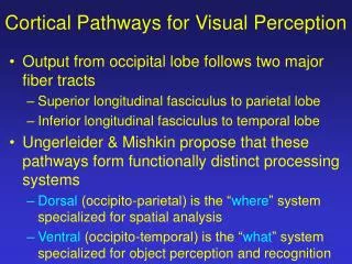
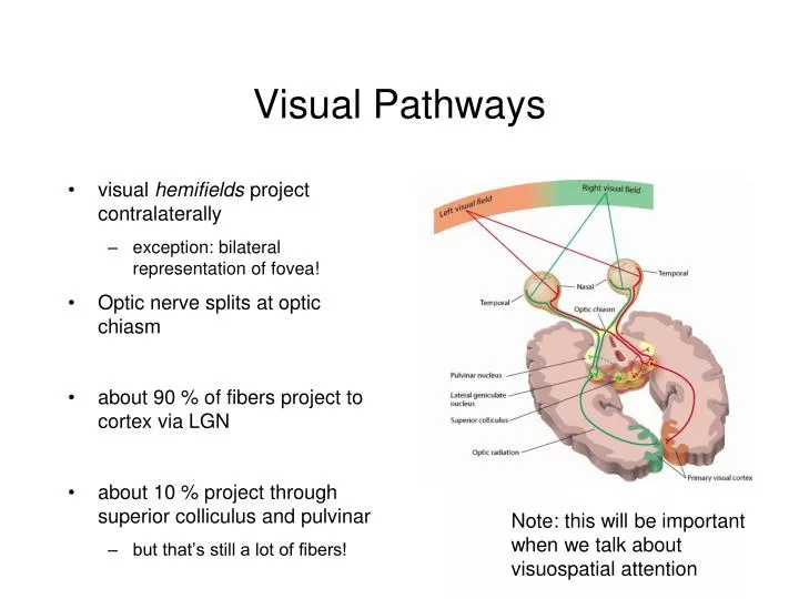
Visual Pathways. visual hemifields project contralaterally exception: bilateral representation of fovea! Optic nerve splits at optic chiasm about 90 % of fibers project to cortex via LGN about 10 % project through superior colliculus and pulvinar but that’s still a lot of fibers! .

E N D
Visual Pathways • visual hemifields project contralaterally • exception: bilateral representation of fovea! • Optic nerve splits at optic chiasm • about 90 % of fibers project to cortex via LGN • about 10 % project through superior colliculus and pulvinar • but that’s still a lot of fibers! Note: this will be important when we talk about visuospatial attention
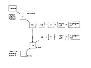
Visual Pathways • Lateral Geniculate Nucleus maintains segregation: • of M and P cells (mango and parvo) • of left and right eyes P cells project to layers 3 - 6 M cells project to layers 1 and 2
Visual Pathways • Primary visual cortex receives input from LGN • also known as “striate” because it appears striped when labeled with some dyes • also known as V1 • also known as Brodmann Area 17
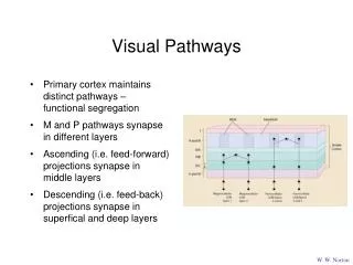
Visual Pathways • Primary cortex maintains distinct pathways – functional segregation • M and P pathways synapse in different layers W. W. Norton
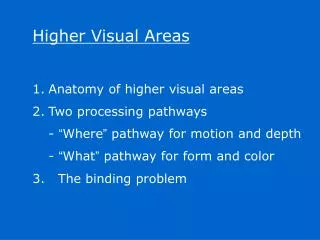
The Role of “Extrastriate” Areas • Different visual cortex regions contain cells with different tuning properties
The Role of “Extrastriate” Areas • Consider two plausible models: • System is hierarchical: • each area performs some elaboration on the input it is given and then passes on that elaboration as input to the next “higher” area • System is analytic and parallel: • different areas elaborate on different features of the input
The Role of “Extrastriate” Areas • Functional imaging (PET) investigations of motion and colour selective visual cortical areas • Zeki et al. • Subtractive Logic • stimulus alternates between two scenes that differ only in the feature of interest (i.e. colour, motion, etc.)
The Role of “Extrastriate” Areas • Identifying colour sensitive regions Subtract Voxel intensities during these scans… …from voxel intensities during these scans …etc. Time ->
The Role of “Extrastriate” Areas • result • voxels are identified that are preferentially selective for colour • these tend to cluster in anterior/inferior occipital lobe
The Role of “Extrastriate” Areas • similar logic was used to find motion-selective areas Subtract Voxel intensities during these scans… …from voxel intensities during these scans …etc. STATIONARY STATIONARY MOVING MOVING Time ->
The Role of “Extrastriate” Areas • result • voxels are identified that are preferentially selective for motion • these tend to cluster in superior/dorsal occipital lobe near TemporoParietal Junction • Akin to Human V5
The Role of “Extrastriate” Areas • Thus PET studies doubly-dissociate colour and motion sensitive regions