Download
1 / 41
410 likes | 593 Vues
Human Anatomy, First Edition McKinley & O'Loughlin. Chapter 23 : Vessels and Circulation. Blood Vessels. An efficient mode of transport for oxygen , nutrients , and waste products to and from body tissues. Heart is the mechanical pump that propels the blood through the vessels.

E N D
Human Anatomy, First EditionMcKinley & O'Loughlin Chapter 23 : Vessels and Circulation
Blood Vessels • An efficient mode of transport for oxygen, nutrients, and waste products to and from body tissues. • Heart is the mechanical pump that propels the blood through the vessels. • Heart and blood vessels form a closed-loop system. • Blood is continuously pumped to and from the tissues. • Are not rigid and immobile. • Can pulsate and change shape in accordance with the body’s needs.
Blood Vessels • Naming: • Often share names with either the body region they traverse or the bone next to them. • Some are named for the structure they supply. • Arteries and veins that travel together sometimes share the same name. • Systemic circulation • consists of the blood vessels that extend to and from the body tissues. • Pulmonary circulation • consists of the vessels that take the blood to the lungs for gas exchange. • Work continuously and intandem with each other.
Three Main Classes of Blood Vessels • Arteries convey blood away from the heart to the body tissues. • Arteries branch, or bifurcate, into smaller and smaller vessels (arterioles) until they feed into the capillaries, where gas and nutrient exchange occurs. • From the capillaries, veins return blood to the heart.
Three Main Classes of Blood Vessels • Arteries become progressively smaller as they divide and get further from the heart. • Veins become progressively larger as they merge and get closer to the heart. • Anastomosis: Site where two or more vessels merge to supply the same body region. • arterial anastomoses: alternate route • Veins tend to form many more anastomoses than do arteries.
Three Main Classes of Blood Vessels • End arteries • Arteries that do not form anastomoses • Only one route • E.g.: renal artery, splenic artery • Functional end arteries • Have small anastomoses • E.g.: coronary arteries
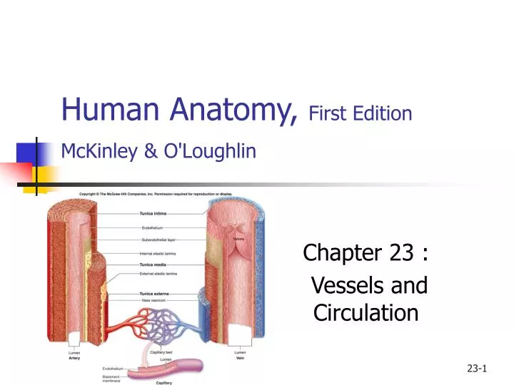
Blood Vessel Tunics • Tunica Intima, or Tunica Interna • innermost layer • composed of: • an endothelium (simple squamous epithelium) • subendothelial layer (areolar CT) • Tunica Media • middle layer of the vessel wall • composed of: • circularly arranged smooth muscle cells • Sympathetic innervation: • Increase: vasoconstriction (narrowing of the blood vessel lumen) • Decrease: vasodilation (widening of the blood vessel lumen)
Blood Vessel Tunics Tunica Externa, or Tunica Adventitia • outermost layer • composed of: • areolar connective tissue that contains elastic and collagen fibers • helps anchor the vessel to other tissues • Term adventitia is used to specify outer layer in blood vessels that are buried in CT • Vasa vasorum : blood vessels that supply large blood vessels • In the externa • Arteries vs Veins: • Media largest in arteries, externa largest in veins • Lumen is smallest in arteries • Artery wall have more elastic and collagen fibers • Capillaries: only the Interna
Arteries • In the systemic circulation, carry oxygenated blood to the body tissues. • Pulmonary arteries carry deoxygenated blood to the lungs. • Three basic types of arteries: • elastic arteries, muscular arteries, and arterioles • as an artery’s diameter decreases • corresponding decrease in the amount of elastic fibers • relative increase in the amount of smooth muscle
Capillaries • Contain only the tunica intima, but this layer consists of a basement membrane and endothelium only. • Allow gas and nutrient exchange between the blood and the body tissues to occur rapidly. • Smallest blood vessels, connect arterioles to venules. • Are called the functional units of the cardiovascular system. • A group of capillaries (10–100) functions together and forms a capillary bed.
The Three Basic Kinds of Capillaries • Continuous capillaries • the most common type • Fenestrated capillaries • Sinusoids, or discontinuous capillaries
Veins • Drain capillaries and return the blood to the heart. • Walls are relatively thin and the vein lumen is larger. • Systemic veins carry deoxygenated blood to the right atrium of the heart, while pulmonary veins carry oxygenated blood to the left atrium of the heart. • Blood pressure is substantially reduced by the time blood reaches the veins. • Hold about 60% of the body’s blood at rest. • Veins function as blood reservoirs.
From Venules to Veins • Venules merge to form veins. • Venule becomes a “vein” when its diameter is greater than 100 micrometers. • Blood pressure in veins is too low to overcome the forces of gravity. • To prevent blood from pooling in the limbs, most veins contain one-way numerous valves to prevent blood backflow in the veins. • As blood flows superiorly in the limbs, the valves close to prevent backflow. • Numerous valves along its length to assist in moving blood back to the heart.
From Venules to Veins Many deep veins pass between skeletal muscle groups. • As the skeletal muscles contract, veins are squeezed to help pump the blood toward the heart. • This process is called the skeletal muscle pump.
Blood Pressure • Force/unit area blood places on the inside wall of a blood vessel. • Measures in mmHg • Sphygmomanometer: device to measure blood pressure. • Systolic blood pressure • Diastolic blood pressure • 120/80 mmHg
Circle of Willis • An important anastomosis of arteries around the sella turcica. • Formed from posterior cerebral arteries and posterior communicating arteries (branches of the posterior cerebral arteries), internal carotid arteries, anterior cerebral arteries, and anterior communicating arteries (which connect the two anterior cerebral arteries). • Equalizes blood pressure in the brain and can provide collateral channels should one vessel become blocked.
Hepatic Portal System • A venous network that drains the GI tract and shunts the blood to the liver for processing and absorption of transported materials. • Blood exits the liver through hepatic veins that merge with the inferior vena cava. • Is needed because the GI tract absorbs digested nutrients, and these nutrients must be processed and/or stored in the liver.
Pulmonary Circulation • Responsible for carrying deoxygenated blood from the right side of the heart to the lungs, and then returning the newly oxygenated blood to the left side of the heart. • Blood is pumped out of the right ventricle into the pulmonary trunk. • This vessel bifurcates into a left pulmonary artery and a right pulmonary artery that go to the lungs.
Aging and the Cardiovascular System • Heart and blood vessels become less resilient. • Elastic arteries are less able to withstand the forces from the pulsating blood. • Systolic blood pressure may increase with age. • Apt to develop an aneurysm, whereby part of the arterial wall thins and balloons out. • Wall is more prone to rupture, which can cause massive bleeding and death. • Incidence and severity of atherosclerosis increases.
Fetal Circulation • Oxygenated blood from the placenta enters through the umbilical vein. • Blood is shunted away from the liver and directly toward the inferior vena cava through the ductus venosus. • Oxygenated blood in the ductus venosus mixes with deoxygenated blood in the inferior vena cava. • Blood empties into the right atrium. • Most of the blood is shunted to the left atrium via the foramen ovale. • Blood flows into the left ventricle and out the aorta. • A small amount of blood enters the right ventricle and pulmonary trunk, but much of this blood is shunted to the aorta through ductus arteriosus. • Blood travels to the rest of the body, and the deoxygenated blood returns to the placenta through umbilical arteries.