Download
1 / 22
240 likes | 505 Vues
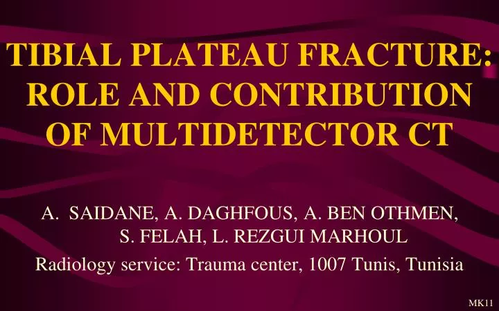
TIBIAL PLATEAU FRACTURE: ROLE AND CONTRIBUTION OF MULTIDETECTOR CT. SAIDANE, A. DAGHFOUS, A. BEN OTHMEN, S. FELAH, L. REZGUI MARHOUL Radiology service: Trauma center, 1007 Tunis, Tunisia. MK11. INTRODUCTION. Tibial plateau fracture occurs mainly in a young population

E N D
TIBIAL PLATEAU FRACTURE: ROLE AND CONTRIBUTION OF MULTIDETECTOR CT • SAIDANE, A. DAGHFOUS, A. BEN OTHMEN, S. FELAH, L. REZGUI MARHOUL Radiology service: Trauma center, 1007 Tunis, Tunisia MK11
INTRODUCTION • Tibial plateau fracture occurs mainly in a young population • It’s consecutive to direct trauma of the knee, generally secondary to traffic accident. • Only explored by the past by plain radiographs, it benefits nowadays of an increasing number of CT exploration. • Our aim is to clarify the role of multidetector scanner in its pretherapeutic assessment.
MATERIAL & METHODS • Retrospectivestudy of 23 patients with fracture of the tibial plateau. • All wereexplored by plain radiographs of the knee and 16 bars CT. • The volume of acquisition ranged from 1 cm above the patella to 1 cm below the tibialtuberosity. • No injection of contrast in the all cases.
MATERIAL & METHODS • Bone and soft tissue filters. • Reconstruction in the coronal and sagittal plans • 3D reconstruction using GE Volume Rendring (VR) • Ten patients were operated • 13 were followed in externe consultation.
RESULTS • Average age = 35 years • Sex ratio (M/W) = 5 • Trauma circumstances: traffic accident (n=9), domestic accident (n=6) and accident at work (n=5) • The tibial plateau fractures were classified according to Duparc et Ficat method
RESULTS • We found: • 9 fractures of the lateral tibial plateau • 5 spino-condylar fractures • 4 medial tibial fractures • 5 bituberosity fractures • More than a third of patients has associated injuries primarily affecting the fibula • 1 case was involved in a polytraumatism
RESULTS (B) (A) Coronal (A) et sagittal (B) reconstructions: Fracture of the lateral plateau with an enfoncement measured at 3.7 mm ( ) and an associated fracture of the lateral femoral condyle ( )
RESULTS 3D reconstructions showing the enfoncement ( ) and the fracture of the femoral condyle ( )
RESULTS (A) (C) (B) Coronal (A), sagittal (B) and 3D (C) reconstructions:comminutive medial spino-condylar fracture
RESULTS (B) Axial (B) and coronal (C) reconstructions: Comminutive fracture of the medial plateau with enfoncement. (A) (A) Coronal reconstruction: Fracture of the medial plateau Separationmeasuredat 5mm (C)
RESULTS Comminutive form of bituberosity fractures
RESULTS Y form of bituberosity fracture T form of bituberosity fracture V form of bituberosity fracture
DISCUSSION • Tibial plateau fractures are secondary to direct trauma on the knee, the more often of a high velocity • Minor trauma may cause similar lesions in case of osteoporosis • Functionnal impotence, pain and knee swelling are the main clinical findings • 2 classifications are used in both plain radiographs and CT
DISCUSSION • The first one, used in France and countries following the french school, was edicted by Duparc and Ficat in 1960 [3,4] and distinguish: • Fractures affecting only one plateau (60%), generally the lateral one, consequently to a direct trauma in valgus (for the lateral plateau) or varus (for the medial) • Squamous tuberosity fracture (10%) • Bituberosity fracture (30%) in V, Y or T. There are also complex and comminutive forms
DISCUSSION • The second classification, anounced by Schatzker [1], divides tibial plateau fractures into 6 types: • Lateral tibial plateau fracture without depression (I) • Lateral tibialplateau fracture withdepression (II) • Compression fracture of the lateral (IIIA) or central (IIIB) tibial plateau • Medial tibial plateau fracture (IV) • Bicondylar tibial plateau fracture (V) • Tibial plateau fracture withdiaphysealdiscontinuity (VI)
DISCUSSION Classification of Schatzker [2]
DISCUSSION • Conflicting data exists regarding the benefit of a pretherapeutic CT scan in these classifications. • Stroet et al[2], Chan et al[5]. et manyothersdid show therewere no increase in agreement between different observers for classification of tibial plateau fractures with the addition of a CT scan comparing to radiographies performed solely. • A possible explanation is that CT provides an overdose of information, which makes classification more difficult.
DISCUSSION • However, practically, CT has many advantages: • Easier classification of fractures, especially between Schatzker I and II [2] • Modification of surgical plans based on plain radiographic findings after CT in 6 to 60% of cases by more preciselydemonstrating the fracture depression and displacementwhich are the most important factors affecting surgical management of standard tibial plateau fractures (NB: 4 mm depression and 2 mm displacementsurgicallevels) [1,4]
DISCUSSION • Diagnosis of associated soft-tissue injuries [1]: According to Gardner et al [6], only 1% of patients with tibial plateau fractures has complete absence of soft-tissue injuries and 77% have cruciate or collateral ligaments lesions that may be suspected in CT Moreover, popliteal vessels lesions (associated with Schatzker IV) are well illustrated by angiographic reconstruction after injection of contrast.
CONCLUSION • Several studies are questioning the superiority of CT in the classification of tibial plateau fractures • However, the scanner offers real practical advantages in the choice of treatment modalities • Besides, the fact that classification methods predate the era of the scanner should promote new methods more more adapted to the scanner
REFERENCES • B. Keegan Markhardt, Jonathan M. Gross, Johnny U. V. Monu, Schatzker Classification of Tibial Plateau Fractures: Use of CT and MR Imaging ImprovesAssessment.RadioGraphics 2009; 29:585–597 2) M. Stroet, M. Holla, J. Biert, A. van Kampen. The value of a CT scan compared to plain radiographs for the classification and treatment plan in tibialplateau fractures. EmergRadiol 2011; 18:279–283 • D. Blin, C. Cyteval, C. Kamba, M. Blondel, FM. Lopez.Imagerie des traumatismes du genou. J Radiol2007; 88: 775-88 • C. Dubois, JN. Ravey, C. Bittighoffer, M. Garelli, T. Delchambre, B. Rubens Duval, N. Mercier, L. Pittet Barbier. TDM et traumatisme des membres inférieurs. JFR 2010 • Chan PSH, Klimkiewicz JJ, Luchetti WT et al (1997) Impact of CT scan on treatment plan and fracture classification of tibialplateau fractures. J Orthop Trauma 11(7):484–489 • Gardner MJ, Geller D, Suk M, et al. The incidence of soft tissue injury in operative tibial plateau fractures: a magnetic resonance imaging analysis of 103 patients. J Orthop Trauma 2005;19(2):79–84