Download
1 / 128
1.35k likes | 1.65k Vues
Chapter 13: The Peripheral Nervous System and Reflex Activity. Peripheral Nervous System (PNS). Links outside world and CNS Includes all neural structures outside the brain and spinal cord Sensory receptors Peripheral nerves and their ganglia Motor endings

E N D
Chapter 13: The Peripheral Nervous System and Reflex Activity
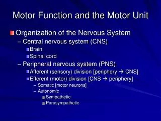
Peripheral Nervous System (PNS) • Links outside world and CNS • Includes all neural structures outside the brain and spinal cord • Sensory receptors • Peripheral nerves and their ganglia • Motor endings • Sensory receptors – respond to changes in environment – stimuli • Activated graded potential nerve impulse • Sensation – awareness of stimuli • Perception – interpretation of meaning • Both occur in brain
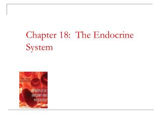
Central nervous system (CNS) Peripheral nervous system (PNS) Sensory (afferent) division Motor (efferent) division Somatic nervous system Autonomic nervous system (ANS) Sympathetic division Parasympathetic division Figure 13.1
Sensory Receptors • Classified according to • Type of stimulus they detect • Body location • Structural complexity
Stimulus Type 1. Mechanoreceptors – mechanical force • Touch pressure (including BP), vibration, and stretch 2. Thermoreceptors– temperature changes 3. Photoreceptors – light energy – retina of eye 4. Chemoreceptors– chemicals in solution • Molecules tasted or smelled, changes in blood or intestinal chemistry 5. Nocieptors– potentially damaging stimuli that result in pain • Searing heat, extreme cold, pressure, inflammatory chemicals
Location 1. Exteroceptors– sensitive to stimuli arising outside of the body • Body surface • Touch, pressure, pain, temperature • Senses – vision, hearing, equilibrium, taste, smell 2. Interoceptors– visceroceptors – stimuli with in the body • Internal viscera and blood vessels • Chemical changes, tissue stretch, temp 3. Proprioceptors– internal stimuli – skeletal muscles, tendons, joints, ligaments, and CT coverings • Advise the brain of body movements
Structural Complexity • Simple Receptors of General Senses – • Receptors respond to several stimuli • 2 types 1. UnencapsualtedDendritic Endings – free or naked nerve endings • Present nearly everywhere • Abundant in CT and epithelia • Unmyelinated, small diameter C fibers • Distal endings – small knoblike swellings • Respond to temperature and painful stimuli
Simple Receptors 1. UnencapsualtedDendritic Endings (cont) • Temperature outside range – cold – 10-40C and hot – 32-48C – perceived as painful • Also respond to pinch and chemicals released by damaged tissue • Itch • Tactile (Merklediscs) – free nerve endings associated with enlarged disc shaped epidermyal cells • Also wrap around hair follicles
Simple Receptors 2. Encapsulated Dendritic Endings – consist of one or more fiber terminals of sensory neurons enclosed in a CT capsule • Most are mechanoreceptors – vary in size, shape, and distribution • Meissner’s corpuscles – small receptors surrounded by Schwann cells and thin CT capsule – touch receptors • Pacinian Corpuscles – lamellated corpuscles – scattered deep in epidermis – pressure • Ruffini Endings – lie in dermis – flattened capsule – deep and continuous pressure
Simple Receptors • 2. Encapsulated Dendritic Endings (cont) – • Muscle spindles – fusiformproprioceptors – perimysium of skeletal muscle – muscle stretch and reflex that resists stretch • Golgi tendon organs – proprioceptors in tendons – tendon fibers stretched – nerve endings are activated • Joint Kinesthetic Receptors – proprioceptors – monitor articular capsules of synovial joints – info on joint position and motion
Complex Receptors • Sense organs • Localized collections of cells associated with the special senses
Sensory Integration • Sensation – awareness of changes in internal and external environment • Perception – conscious interpretation of these stimuli • We depend on both to survive
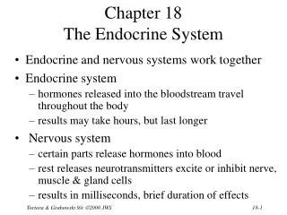
Somatosensory System • Part of the sensory system serving the body wall and limbs • Receives input from exteroceptors, proprioceptors, and interoceptors • 3 main levels of neural integration – • 1. receptor level – sensory receptors • 2. Circuit level – ascending pathways • 3. Perceptual level – neuronal circuits in cerebral cortex
Perceptual level(processing in cortical sensory centers) 3 Motor cortex Somatosensory cortex Thalamus Reticular formation Cerebellum Pons Medulla Circuit level (processing in ascending pathways) 2 Spinal cord Free nerve endings (pain, cold, warmth) Muscle spindle Receptor level (sensory reception and transmission to CNS) 1 Joint kinesthetic receptor Figure 13.2
1. Receptor Level • Sensation – stimulus must excite a receptor and APs must reach the CNS • For this to happen – stimulus – • energy must match specifically to receptor • must be applied within the receptive field • Energy must be converted into a graded potential (receptor potential) by transduction • Generator potential in the associated neuron must reach a threshold
1. Receptor Level • Adaptation– sensory receptors can change sensitivity in presence of a constant stimulus • Phasic receptors – fast adapting – bursts of impulses at the begging and end of stimulus • Tonic Receptors – sustained response – little or no adaptation
2. Circuit Level • Delivers impulses to the cerebral cortex for stimulus localization and perception
3. Perceptual Level • Interpretation of Sensory input in cerebral cortex • Projection – exact point in cortex that is activated is always the same “where” regardless of how it is activated
3. Perceptual Level • Sensory Perception – • Perceptual detection – ability to detect that a stimulus has occurred • Magnitude Estimation – ability to detect how intense the stimulus is • Spatial discrimination – identify the site or pattern of stimulation • Feature Abstraction – mechanism by which one neuron or circuit is turned to one feature in the presence of another • Quality discrimination – ability to differentiate submodalities (qualities) of a sensation • Pattern recognition – ability to take in the scene around us and recognize a familiar pattern
Perception of Pain • Receptors activated by extremes of pressure and temperature, as well as, chemicals release by damaged tissue • Sharp pain – small myelinated A delta fibers • Burning pain – small unmyelinated C fibers • Both release glutamate and substance P activate 2nd order neurons • Hyperalgesia – pain amplification • Phantom Limb pain – pain in tissue that is no longer present
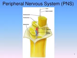
Transmission Lines – Nerves & Their Ganglia • Structure and Classification – • Nerve – cordlike organ • Vary in size • Consists of parallel bundles of peripheral axons enclosed by CT • Axon – surrounded by endoneurium – CT layer • Groups of fibers (fascicles) bound together by perineurium • Finally fascicles are enclosed by - epineurium
Axon Myelin sheath Endoneurium Perineurium Epineurium Fascicle Blood vessels (b) Figure 13.3b
Nerves & Their Ganglia • Classified according to the direction which they transmit impulses • Mixed nerves – both ways • Sensory (afferent) nerves – carry impulses towards the CNS • Motor (efferent) nerves – carry impulses away from CNS • Ganglia – collections of neuron cell bodies associated with nerves in the PNS
Regeneration of Nerve Fibers • Real mature neurons do not divide • Damage severe or close to cell body – entire neuron may die • Other neurons attached to that neuron may also die • Cell body intact – cut or compressed nerves can regenerate successfully
Regeneration of Nerve Fibers • Axon becomes fragmented at the injury site • Macrophages clean out the dead axon distal to injury • Axon sprouts, or filaments, grow through a regeneration tube formed by Schwann cells • The axon regenerated and a new myelin sheath forms
Endoneurium Schwann cells 1 The axon becomes fragmented at the injury site. Droplets of myelin Fragmented axon Site of nerve damage Figure 13.4 (1 of 4)
Macrophages clean out the dead axon distal to the injury. 2 Schwann cell Macrophage Figure 13.4 (2 of 4)
Axon sprouts, or filaments, grow through a regeneration tube formed by Schwann cells. 3 Aligning Schwann cells form regeneration tube Fine axon sprouts or filaments Figure 13.4 (3 of 4)
The axon regenerates and a new myelin sheath forms. 4 Site of new myelin sheath formation Schwann cell Single enlarging axon filament Figure 13.4 (4 of 4)
Cranial Nerves • 12 pairs associated with brain • 1st – forebrain • Rest – brain stem • Only head and neck structures
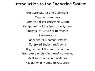
Filaments of olfactory nerve (I) Frontal lobe Olfactory bulb Olfactory tract Optic nerve (II) Temporal lobe Optic chiasma Infundibulum Optic tract Facial nerve (VII) Oculomotor nerve (III) Trochlear nerve (IV) Vestibulo- cochlear nerve (VIII) Trigeminal nerve (V) Glossopharyngeal nerve (IX) Abducens nerve (VI) Vagus nerve (X) Cerebellum Accessory nerve (XI) Medulla oblongata Hypoglossal nerve (XII) (a) Figure 13.5 (a)
Cranial nerves I – VI Sensory function Motor function PS* fibers I Olfactory Yes (smell) No No II Optic Yes (vision) No No III Oculomotor No Yes Yes IV Trochlear No Yes No V Trigeminal Yes (general sensation) Yes No VI Abducens No Yes No Cranial nerves VII – XII Sensory function Motor function PS* fibers VII Facial Yes (taste) Yes Yes VIII Vestibulocochlear Yes (hearing and balance) Some No IX Glossopharyngeal Yes (taste) Yes Yes X Vagus Yes (taste) Yes Yes XI Accessory No Yes No XII Hypoglossal No Yes No *PS = parasympathetic (b) Figure 13.5 (b)
Cranial Nerves I. Olfactory – tiny sensory nerves of smell • Run from nasal mucosa to synapse with the olfactory bulb II. Optic – sensory nerve of vision – brain tract III. Oculomotor– “eye mover” – 6 extrinsic muscles that move the eye IV. Trochlear– “pulley” innervates extrinsic eye muscle through a pully shaped ligament
Cranial Nerves V. Trigeminal – 3 branches, sensory fibers to the face and motor fibers to the chewing muscles VI. Abducens– controls extrinsic eye muscle that abducts the eyeball VII. Facial – large nerve – innervates muscles of facial expression VIII. Vestibulocochlear– auditory nerve – hearing and balance
Cranial Nerves IX. Glossopharyngeal– tongue and Pharynx X. Vagus– only cranial nerve that extends beyond the head into the thorax and abdomen XI. Accessory – accessory part of the vagus nerve XII. Hypoglossal – under the tongue, innervates the tongue muscles