Download
1 / 64
650 likes | 1.01k Vues
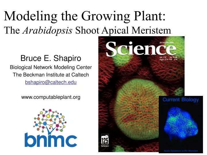
Modeling the Growing Plant: The Arabidopsis Shoot Apical Meristem. Bruce E. Shapiro Biological Network Modeling Center The Beckman Institute at Caltech bshapiro@caltech.edu www.computableplant.org. Some Background Information Fluorescent Probe Confocal Microscopy Arabidopsis

E N D
Modeling the Growing Plant: The Arabidopsis Shoot Apical Meristem Bruce E. Shapiro Biological Network Modeling Center The Beckman Institute at Caltech bshapiro@caltech.edu www.computableplant.org
Some Background Information • Fluorescent Probe Confocal Microscopy • Arabidopsis • Connectivity & Cell Walls form Delaunay and Voronoi • Cell Division and Plant Growth • Phyllotaxis • Cell Differentiation
http://www.computableplant.org • National Science Foundation (US) Frontiers in Integrative Biological Research (FIBR) Program Grants (First grants: Sept. 2003) • must integrate research tools from across multiple disciplines: biology, math, physical sciences, information technology • not limited by organizational boundaries • must answer a fundamental biological question • 5 year duration, renewable, ≈$1,000,000/year • 5± new grants annually
"To analyze the charms of flowers is like dissecting music; it is one of those things which it is far better to enjoy than to attempt to understand.” [Henry Tuckerman, 1853] • “The project will track cell-by-cell changes in the mustard plant's meristem - the tissue in which cells actively divide and then differentiate into specialized cells. With fluorescent proteins marking specific cell types in specially designed transgenic plants, researchers will be able to trace the development of leaves and flowers.” [NSF Press Release, 2003]
Eric Mjolsness (PI) Pierre Baldi Alex Sadovsky Tigran Bacarian Alexi Vorbyov Ashish Bhan Fang Fang Elliot Meyerowitz Marcus Heisler Venu Reddy Adrienne Roeder Vikas Agrawal* Bruce Shapiro Victoria Gor Nikolai Kolchanov Nadya Omelianchuk Nikolay Podkolodny Sergei Nikolaev Vitali Likhoshvai Henrik Jönsson US National Science Foundation FIBR Grant 0330786
... to bring together Caltech biologists, bioengineers, mathematicians, and computer scientists to develop and apply state-of-the-art computational tools for modeling and analyzing complex biological systems.
Mjolsness E. Stochastic Process Semantics for Dynamical Grammer Syntax: An Overview. 9th International Symposium on Artificial Intelligence and Mathematics, Jan 2006. Reddy GV, Meyerowitz EM. Stem-Cell Homeostasis Growth Dynamics can be Uncoupled in the Arabidopsis Shoot Apex. Science 310(5748):663-667.
Systems Biology Ontology
Fluorescent Probe Confocal Microscopy • Arabidopsis • Connectivity & Cell Walls form Delaunay and Voronoi • Cell Division and Plant Growth • Phyllotaxis • Cell Differentiation
lifesci.ucsb.edu/~biolum www.reefs.org Luminescent Jellyfish Aequoria victoria
Marc Zimmer (Photo: Osamu Shimomura) http://www.conncoll.edu/ccacad/zimmer/GFP-ww/GFP2.htm Luminescent Jellyfish Aequoria victoria
Marc Zimmer GFP: Green Fluorescent Protein
Commercial Markers www.anticancer.com Neurons J. Lichtman,Science (2003) Transgenic Art Eduardo Kac Nikon’s Microscopyu.com GFP: Green Fluorescent Protein
Fluorescence Microscopy Higher Energy State Internal Energy Loss Intermediate State Sample absorbs high energy photon from laser Photon Emitted Ground State
Laser Scanning Confocal MicroscopeZeiss LSM-510 Venu Reddy Marcus Heisler
Fluorescent Probe Confocal Microscopy • Arabidopsis • Connectivity & Cell Walls form Delaunay and Voronoi • Cell Division and Plant Growth • Phyllotaxis • Cell Differentiation
Geographical distribution of Arabidopsis thaliana. Koornneff, Ann. Rev. Plant Biol. 2004
Cell 88:299-308 (1997) Live Imaging
51 image “z-stack”
PIN1:GFP (blue),REVOLUTA:YFP (green)andpFIL:dsRED (red) Marcus Heisler, Caltech
Cell 88:299-308 (1997) Tracing Cell Lineages Development 131:4225 (2004) Reddy et al
Tracking cell motion Thin Plate Splines Softassign
Auxin Derived from PIN1 pH=5.0 pH=4.5 pH=5.5
Fluorescent Probe Confocal Microscopy • Arabidopsis • Connectivity & Cell Walls form Delaunay and Voronoi • Cell Division and Plant Growth • Phyllotaxis • Cell Differentiation
DiscreteMath`ComputationalGeometry`DelaunayTriangulation qdelaunay regtet
Fluorescent Probe Confocal Microscopy • Arabidopsis • Connectivity & Cell Walls form Delaunay and Voronoi • Cell Division and Plant Growth • Phyllotaxis • Cell Differentiation
Shoot Apical Meristem - tip of plant Central Zone: Provides new cells Flower bud Peripheral Zone: Leaves, Flower Formation MERISTEM Rib:Cells divide and “push” Meristem up STEM 50 scale bar
Protein regulation model Internal External Receptor, c - Ligand, b Mjolsness, Sharp & Reinitz J. Theor. Biol. 152: 429 (1991) Shapiro, Levchenko, Mjolsness, Meyerowitz, Wold, Bioinformatics19(5):677 (2003)
Growth Model • Constant growth rate: • Cell division on mass threshold • Viscous spring model:
Meristem Maintenance and Growth Mjolsness, Shapiro, Jönnson & Meyerowitz in BGRS, Kluwer (2002); also EMBS (2002)
Fluorescent Probe Confocal Microscopy • Arabidopsis • Connectivity & Cell Walls form Delaunay and Voronoi • Cell Division and Plant Growth • Phyllotaxis • Cell Differentiation
Auxin - A Plant Hormone Indole-3-acetic acid Synthesized from tryptophan?

![Data Modeling [Comparison of data modeling techniques ]](https://cdn0.slideserve.com/205866/data-modeling-comparison-of-data-modeling-techniques-dt.jpg)