Download
1 / 67
710 likes | 754 Vues
ANATOMY OF TRACHEOBRONCHIAL TREE AND PULMONARY VENTILATION Presented by Dr. Sunil Kumar Arora. Organs of the Respiratory System A. The organs of the respiratory tract can be divided into two groups: 1.Upper Airways 2.Lower airways. Upper Airways. Nose Paranasal sinuses

E N D
ANATOMY OF TRACHEOBRONCHIAL TREE AND PULMONARY VENTILATION Presented by Dr. Sunil Kumar Arora
Organs of the Respiratory System A. The organs of the respiratory tract can be divided into two groups: 1.Upper Airways 2.Lower airways
Upper Airways Nose Paranasal sinuses Pharynx
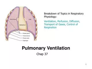
Larynx Trachea Main stem bronchi Segmental bronchi Subsegmental bronchi Bronchioles Terminal bronchioles Respiratory bronchioles Alveolar ducts Alveolar sacs alveoli Lower AirwayBegins with true vocal cords and extends to alveoli
Organization and Functions of the Respiratory System • Conducting portion transports air. - includes the nose, nasal cavity, pharynx, larynx, trachea, and progressively smaller airways, from the primary bronchi to the terminal bronchioles • Respiratory portion carries out gas exchange. - composed of small airways called respiratory bronchioles and alveolar ducts as well as air sacs called alveoli
Tracheobronchial Tree • A highly branched system of air-conducting passages that originate from the trachea and progressively branch into narrower tubes as they diverge throughout the lungs before terminating in terminal bronchioles. • Series branching airways commonly referred to as “generations” or of “orders” The first generation or order is zero (0), the trachea itself
Trachea • A flexible tube of 10-12 cm length and 1.5-2.5 cm width also called windpipe. Extends to second rib anteriorly and T4-T5 posteriorly. • Extends through the mediastinum and lies anterior to the esophagus and inferior to the larynx. • Anterior and lateral walls of the trachea supported by 15 to 20 C-shaped tracheal cartilages. • Cartilage rings reinforce and provide rigidity to the tracheal wall to ensure that the trachea remains open at all times • Posterior part of tube lined by trachealis muscle.
Tracheal lining • Pseudostratified columnar epithelium with cilia; goblet cells, serous cells, and specialized submucosal bronchial glands • 200+ cilia per cell, 5-7 microns long • Beat cephalid (head) toward oropharynx
Primary bronchus • At the level of the sternal angle, the trachea bifurcates into two smaller tubes, called the right and left primary bronchi. • Each primary bronchus projects laterally toward each lung. • The most inferior tracheal cartilage separates the primary bronchi at their origin and forms an internal ridge called the carina.
Right bronchus Wider More vertical 5 cm shorter Supported by C shaped cartilages 20-30 degree angle First generation Left bronchus Narrower More angular Longer Supported by C shaped cartilages 40-60 degree angle First generation Main Stem Bronchi • Foreign particles are more likely to lodge in the right primary bronchus.
Bronchial tree • The primary bronchi enter the hilus of each lung together with the pulmonary vessels, lymphatic vessels, and nerves. • Each primary bronchus branches into several secondary bronchi (or lobar bronchi). • The left lung has two secondary bronchi.The right lung has three secondary bronchi. • They further divide into tertiary bronchi. • Each tertiary bronchus is called a segmental bronchus because it supplies a part of the lung called a bronchopulmonary segment.
R main stem divides into: Upper lobar bronchus Middle lobar bronchus Lower lobar bronchus L main stem divides into: Upper lobar bronchus Lower lobar bronchus Lobar Bronchi
R lobar divides into Segmental bronchi 10 segments on right L lobar divides into Segmental bronchi 8 segments on left Segmental Bronchi 3rd generation
Subsegmental Bronchi • 4th to 9th generations • Progressively smaller airways • 1-4 mm diameter • At 1 mm diameter connective tissue sheath disappears
10-th to 15th generation Cartilage is absent Lamina propria is directly connected with lung parenchyma Surrounded by spiral muscle fibers Epithelial cells are cuboidal Less goblet cells and cilia With no cartilage, airway remains open due to pressure gradients Noncartilagenous Airways Bronchioles
Terminal Bronchioles • 16th to 19th generation • Average diameter is 0.5 mm • Cilia and mucous glands begin to disappear totally • End of the conducting airway • Canals of Lambert-interconnect this generation,provide collateral ventilation
Respiratory Bronchioles, Alveolar Ducts, and Alveoli • Lungs contain small saccular outpocketings called alveoli. • They have a thin wall specialized to promote diffusion of gases between the alveolus and the blood in the pulmonary capillaries. • Gas exchange can take place in the respiratory bronchioles and alveolar ducts as well as in the alveoli, each lung contains approximately 300 to 400 million alveoli. • The spongy nature of the lung is due to the packing of millions of alveoli together.
Gas exchange zone • Respiratory bronchioles • Acinus • Alveolar Ducts, sacs, alveoli
Functional Units of Gas Exchange • Three generations of respiratory bronchioles • Three generations of alveolar ducts • 15-20 clusters--sacs
Acinus or Lobule • Each acinus (unit) is approximately 3.5 mm in diameter • Each contains about 2000 aveloli • There are approximately 130,000 primary lobules in the lung
Anatomic Arrangement of Alveoli • 85-95% of alveoli covered by small pulmonary capillaires • The cross-sectional area or surface area is approximately 70m2 • Total of about 300 million alveoli in 2 lungs
Cells in Alveolus Type I cells : 95% of Alveoler surface, Major site of gas exchange Type II cells : or Septal cells secrete surfactant,About 5% of Alveoler surface. Alveolar macrophages-Plays role in defence mechenism.
Surfactant • Is a substance produce by type II alveolar epithelial cells (~ 5% of the surface area of the alveoli) which reduce the surface tension of the fluid in the inner surface of the alveoli • it is a mixture of phospholipids, proteins, and ions, the most important component is phospholipid dipalmitoyl phosphatidylcholine which is responsible for reducing the surface tension.
Respiratory Membrane Diffusion of gases occurs through Respiratory mambrane. • squamous cells of alveoli . • basement membrane of alveoli. • basement membrane of capillaries • simple squamous cells of capillaries • about .5 μ in thickness
Epithelial basement membrane Capillary basement membrane Interstitial space Capillary endothelium Alveolar epithelium Red blood cell Fluid and surfactant layer Alveolus Capillary Diffusion O2 Diffusion CO2
Intersitium/interstial space • Surround, supports, and shapes the alveoli and capillaries • Composed of a gel like substance and collagen fibers • Contains tight space and loose space areas
Factors that affect the rate of gas diffusion through the respiratory membrane • The thickness of the respiratory membrane.The thickness of the respiratory membrane is inversely proportional to the rate of diffusion through the membrane. • Surface area of the membrane.
The diffusion rate of the specific gas. Diffusion coefficient for the transfer of each gas through the respiratory membrane depends on its solubility in the membrane and inversely on the square root of its molecular weight. CO2 diffuses 20 times as rapidly as O2. • The pressure difference between the two sides of the membrane (between the alveoli and in the blood).
Pores of Kohn • Small holes in the walls of adjoining alveoli (alveaolar septa) • Between 3 to 13 µ in diameter • Formation of pores may be due to: • Desquamation due to disease • Normal degeneration due to aging • Movement of macrophages leaving holes
Blood supply of Lungs • Bronchial circulation – bronchial arteries supply oxygenated blood to lungs, bronchial veins carry away deoxygenated blood from lung tissue. • Pulmonary circulation • Response of two systems to hypoxia – pulmonary vessels undergo vasoconstriction, bronchial vessels like all other systemic vessels undergo vasodilation
Bronchial Blood Supply Bronchial arteries Arise from thoracic aorta(Anatomic variation is there),usually there are 1 artery for Rt lung and 2 arteries for Lt Lung. Bronchial arteries run along with the branching bronchi and supply lung tissue except the alveoli. Much of the blood supplied by Bronchial arteries is Returned via Pulmonary veins rather than bronchial veins.
Bronchial arteries • Also nourish • Mediastinal lymph nodes • Pulmonary nerves • Some muscular pulmonary arteries and veins • Portions of the esophagus • Visceral pleura
Bronchial venous system • Approx.1/3 blood returns to right heart • Azygous vien • Hemiazygous vein • Intercostal veins • This blood comes form the first two or three generations of bronchi
Bronchial venous return • 2/3 of blood flowing to terminal bronchioles drains into pulmonary circulation via “bronchopulmonary anastomoses” • Then flows to left atrium via pulmonary veins • Contributes to “venous admixture” or “anatomic shunt” (1-2% of C.O.)
Pulmonary Vascular System • The second source of blood to the lungs • Primary purpose is to deliver blood to lungs for gas exchange • Also delivers nutrients to cells distal to terminal bronchioles • Composed of arteries, arterioles, capllaries, venules, and veins
Pulmonary Trunk arises from the infundibulum of Rt ventricle and divide into Rt and Lt branches.Which carry deoxygenated blood. • Pulmonary arteries branch profusely along with the bronchi leading to capillary network sorrunding the alveoli. • 4 Pulmonary veins(2 from each Lung) form post alveoli to carry oxygenated blood to the Lt Atrium
Pulmonary Capillaries • Walls are less than 0.1µ thick • Total external thickness is about 10µ • Selective permeability to water, electrolytes, sugars • Produce and destroy biologically active substances
Nerve Supply • Lung and Pleura are innervated by Anterior and posterior pulmonary plexuses • They are mixed plexuses containing vagal(Parasympethatic) and sympathetic fibers. • Vagus Nerve(Parasympathetic)—Bronchoconstriction,vasodilatation and Secretomotor • Sympathetic Fibres—Bronchodilator,vasocostrictor and inhibitor to the glands of bronchial tree.
Lymphatic System Lymphatic vessels remove fluids and protein molecules that leak out of the pulmonary capillaries Transfer fluids back into the circulatory system
Lymphatic Drainage of Lungs • There are 2 sets of Lymphatics,both of which drain into the Bronchopulmonary nodes. • Superficial vessels drain the peripheral lung tissue lying beneth the pulmonary pleura • Deep lymphatics drain the bronchial tree,the pulmonary vessels and connective tissue septa and then run towards the hilum.
Lymphatic vessels of upper lobe end up in superior tracheobronchial node and from lower lobe enters the inferior tracheobronchial nodes. • The tracheobronchial and bronchopulmonary nodes end in the bronchomediastinal lymph trunk • This trunk reaches the thoracic duct on the left and Rt lymphatic duct on the Rt side.
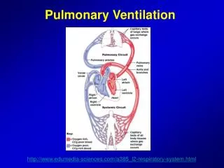
Respiratory events • Pulmonary ventilation = exchange of gases between lungs and atmosphere • External respiration = exchange of gases between alveoli and pulmonary capillaries • Internal respiration = exchange of gases between systemic capillaries and tissue cells
Breathing • Breathing (pulmonary ventilation). consists of two cyclic phases: • inhalation, also called inspiration - draws gases into the lungs. • exhalation, also called expiration - forces gases out of the lungs.
Muscles of Inspiration • During inspiration, the dome shaped diaphragm flattens as it contracts • This increases the height of the thoracic cavity • The external intercostalmuscles contract to raise the ribs • This increases the circumference of the thoracic cavity Together: