Download
1 / 123
1.33k likes | 1.98k Vues
CPAP for the Wisconsin EMT. Continuous Positive Airway Pressure. Objectives. Introduction to CPAP Review Pulmonary Anatomy and Physiology Review pathophysiology of respiratory distress CPAP Delivery Systems Protocol for CPAP administration Required data collection. Goal of CPAP.

E N D
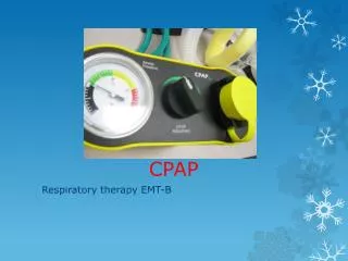
CPAP for the Wisconsin EMT Continuous Positive Airway Pressure
Objectives • Introduction to CPAP • Review Pulmonary Anatomy and Physiology • Review pathophysiology of respiratory distress • CPAP Delivery Systems • Protocol for CPAP administration • Required data collection
Goal of CPAP To have an effective way to treat respiratory distress from COPD, Asthma, CHF, and pneumonia without having to intubate the patient • Provide “splinting” of the airways • Enhance gas exchange • Provide a more effective delivery of medications • Albuterol/Atrovent
History of CPAP 1912 - Maintenance of Lung Expansion during Thoracic Surgery (S. Brunnel) 1937 - High Altitude flying to prevent hypoxemia. (Barach et al) 1967 - CPPB + IPPV to treat ARDS (Ashbaugh et al) 1971 - Term CPAP introduced, used to treat HMD in neonates (Gregory et al) 1972 - CPAP used to treat ARF (Civetta et al) 1973 - CPAP used to treat COPD (Barach et al) 1981 - Downs generator (Fried et al) 1982 - Modern definition of CPAP (Kielty et al) Theory has been around a long time!
Continuous Positive Airway Pressure How it Works Flow Generator Produces pressure by using an oxygen source A venturi effect entrains ambient air
Continuous Positive Airway Pressure How it Works Positive End-Expiratory Pressure (PEEP) Valve The peep valve maintains a set cm H2O pressure
Continuous Positive Airway Pressure How it Works With each breath Oxygen-enriched air is forced and maintained in the patient’s airway and lungs and Fluid is forced out of the alveolar space Occurs at 110-115 liters / min.
CPAP and Patient Airway Pressure ‘The application of positive airway pressure throughout the whole respiratory cycle to spontaneously breathing patients’
Continuous Positive Airway Pressure BiPAP versus CPAP BiPAP: Provides a biphasic pressure by decreasing the pressure during exhalation CPAP: Provides continuous pressure throughout respiratory cycle BiPAP results in lower work of breathing. Unfortunately, no pre-hospital BiPAP device exists.
Airway Anatomy and Physiology • Respiratory structures are divided by their locations relative to the glottic opening or vocal cords • Upper airway - structures located above glottis • Lower airway - structures located below glottis
UPPER AIRWAY Nares Nasopharynx Oropharynx Tongue Epiglottis/Glottis Vocal Cords LOWER AIRWAY Trachea/Esophagus Carina Main stem Bronchi Secondary Bronchi Bronchioles Alveoli Elements of the Airway
Pharynx • Nasopharynx • Uppermost portion of airway, just behind nasal cavities • Nasal septum • Vestibule • Olfactory membranes • Sinuses • Oropharynx • Begins at the level of the uvula and extends down to the epiglottis • Opens into the oral cavity
Larynx • Three main functions: • Air passageway between the pharynx and lungs • Prevents solids and liquids from entering the respiratory tree • Involved in speech production
Larynx • An outer casing of nine cartilages • Thyroid cartilage • Cricoid cartilage • Only complete cartilaginous ring in the larynx • Epiglottis • Hyoid bone • Cricothyroid membrane • Vocal cords
Lower Airway Structures • Trachea • Bronchial tree • Primary bronchi • Secondary bronchi • Bronchioles
Lower Airway Structures • Alveoli
Lungs • Principal function is respiration • Attached to heart by pulmonary arteries and veins • Separated by mediastinum and its contents • Base of each lung rests on the diaphragm • Apex extends 2.5 cm above each clavicle
Pleural Cavity • A separate pleural cavity surrounds each lung • Two layers (visceral and parietal) • Pleural space
Respiratory System - Physiology • The respiratory system functions as a gas exchange system • Oxygen is diffused into the bloodstream for use in cellular metabolism
Respiratory System - Physiology • Wastes, including carbon dioxide, are excreted from the body via the respiratory system
Pathophysiology • Specific pathophysiologies responsible for respiratory emergencies include those related to ventilation, diffusion, and perfusion
Ventilation • Ventilation refers to the process of air movement in and out of the lungs • The volume of air moved in each breath is the tidal volume • The volume still remaining in the chest after exhalation is the functional reserve capacity. FRC
Inspiration and Expiration • Inspiration • Chest wall expands • Lung space increases • Pressure gradient causes gas to flow into the lungs • Expiration • Chest wall relaxes • Elastic recoil causes thorax and lung space to decrease in size • Pressure gradient created in thoracic cavity causes air to move out of the chest
Pressure Changes During Inspiration and Expiration Figure 11-1
Ventilation • The following must be intact for ventilation to occur: • Neurologic control to initiate ventilation • Nerves between the brainstem and the muscles of respiration • Functional diaphragm and intercostal muscles • A patent upper airway • A functional lower airway • Alveoli that are functional and not collapsed
Diffusion • In order for diffusion to occur, the following must be intact: • Alveolar and capillary walls that are not thickened • Interstitial space between the alveoli and capillary wall that is not enlarged or filled with fluid
Respiratory Vocabulary • Dyspnea –Difficult or labored breathing • Hypoxia-State in which insufficient oxygen is available to meet the oxygen requirement of the cells • Hypercarbia-Build up of carbon dioxide • Asphyxia-Interference with respiration producing hypoxia, and hypercarbia
More Terms • Nasal Flaring-Excessive widening of the nares with respiration • Cyanosis-Bluish discoloration of the skin due to hypoxia • Snoring-Occurs when the upper airway is partially obstructed • Stridor-Harsh, high pitched sound heard on inspiration. Secondary to upper airway obstruction such as croup
Even more terms • Wheezing-Whistling sound, usually on expiration, and due to narrowing of the lower airway • Rhonchi-Musical/rattling in the lower airways, usually from excessive mucous • Rales-Crackling sounds associated with fluid around the alveoli. Can occur with CHF or Pneumonia • Accessory muscle use and retractions – the use of chest muscles to expand the chest and the “sucking” of the skin between the boney skeleton of the chest during inspiration
The Physiology of CPAP CPAP will eventually decrease the work of breathing by improving the hypoxic state: 1. Forces fluid out of the alveolar space back into the interstitium 2. Forces oxygen-enriched air into the lungs to improve diffusion and uptake on hemoglobin
Continuous Positive Airway Pressure Pressure
CPAP and Partial Pressure ‘The pressure of a gas mixture is equal to the sum of the partial pressures of its constituents’ This allows oxygen into the blood during inspiration and Carbon Dioxide out during expiration. Example : Air at sea level has a pressure of 760mm Hg Air is 21% oxygen and 79% nitrogen partial pressure of oxygen is 760 X 21% = 159mm Hg
Pressure Gradient Deoxygenated blood has a lower partial pressure of oxygen than alveolar air so oxygen transfers from the air into the blood
Pressure Gradient 5.0 cm H20 CPAP 1cm H2O is equal to 0.735mm Hg. 1.0 cm H2O CPAP increases the partial pressure of the alveolar air by approximately 1%. This increase in partial pressure ‘forces’ more oxygen into the blood. Even this comparatively small change is enough to make a clinical difference.
The Requirements Of CPAP • The real requirement is for Continuous CONSTANT Positive Airways Pressure • A stable airway pressure as prescribed in order to reduce work of breathing (WOB)
Important Aim Of CPAP Is To Increase Functional Residual Capacity (FRC) • Volume of gas remaining in lungs at end-expiration • CPAP distends alveoli preventing collapse on expiration • Greater surface area improves gas exchange
Physiological Effects Of CPAP • Increases PSO2 • Increases FRC • Reduces Work of Breathing
CPAP And Pulmonary Edema • Severe Pulmonary Edema is a frequent cause of respiratory failure • CPAP increases functional residual capacity • CPAP increases transpulmonary pressure • CPAP improves lung compliance • CPAP improves arterial blood oxygenation • CPAP redistributes extravascular lung water (Rasanen 1985)
CPAP And Acute Respiratory Failure • CPAP overcomes inspiratory work imposed by auto-peep • CPAP prevents airway collapse during exhalation • CPAP improves arterial blood gas values • CPAP may avoid intubation and mechanical ventilation (Miro 1993)
Congestive Heart Failure and Pulmonary Edema With CHF and Pulmonary Edema: • Fluid backs up into pulmonary veins • Increase in hydrostatic pressure • Fluid traverses the capillary membrane • Fluid enters the alveolar spaces
Congestive Heart Failure and Pulmonary Edema • With CHF:
Congestive Heart Failure and Pulmonary Edema • With CHF:
Congestive Heart Failure and Pulmonary Edema • With CHF: