Download
1 / 91
1.03k likes | 1.34k Vues
Emergency Ultrasound in Trauma. Anthony J Weekes MD, RDMS Janet G. Alteveer, MD Sarah Stahmer, MD. Clinical Case.

E N D
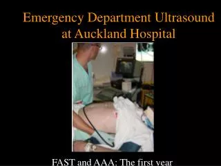
Emergency Ultrasound in Trauma Anthony J Weekes MD, RDMS Janet G. Alteveer, MD Sarah Stahmer, MD
Clinical Case • GR is a 62 y male who hit his right torso when he slipped on an icy sidewalk. He denies head trauma, and can walk without a limp. Two hours later the pain in his lower chest has increased he comes to the ED.
Clinical Case • PE: BP116/72, pulse109, RR 24. • There is a minor abrasion to right lateral chest, which is tender to palpation. Diffuse mild abdominal tenderness. • Meds: Coumadin for irregular heartbeat
Clinical Case • 2 large IV’s placed, CXR done. Blood tests sent. • Bedside ultrasound done. • CXR revealed lower rib fractures, no HTX or PTX
Clinical Case • FFP ordered and OR notified. • He is found to have a liver laceration and 500 cc of blood in the peritoneal cavity.
Diagnostic Modalities in Blunt Abdominal Trauma • Diagnostic Peritoneal Lavage (DPL) • CAT Scan • Ultrasound (FAST exam)
Advantages Very sensitive for identifying intra-peritoneal blood 106 RBC/mm3 approx. 20 ml blood in 1L lavage fluid Can be done at the bedside Can be done in 10-15 minutes Disadvantages Overly sensitive, may result in too high a laparotomy rate Invasive Difficult in pregnancy, or with many prior surgeries Can not be repeated Diagnostic Peritoneal Lavage
Advantages Identifies specific injuries Good for hollow viscus and retroperitoneal injury High sensitivity and specificity Disadvantages Expensive equipment 30-60 minutes to complete study Only for stable patients Not for pregnant patients CT Scan
Advantages Can be performed in 5 minutes at the bedside Non-invasive Repeat exams Sensitivity and specificity for free fluid equal to DPL and CT Disadvantages Operator dependent May not identify specific injury Poor for hollow viscus or retroperitoneal injury Obesity, subcutaneous air may interfere with exam FAST
FAST Principles • Detects free intraperitoneal fluid • Blood/fluid pools in dependent areas • Pelvis • Most dependent • Hepatorenal fossa • Most dependent area in supramesocolic region
FAST Principles • Pelvis and Supra-mesocolic areas communicate • Phrenicolic ligament prevents flow • Liver/spleen injury • Represents 2/3 of cases of blunt abdominal trauma
FAST- principles • Intraperitoneal fluid may be • Blood • Preexisting ascites • Urine • Intestinal contents
FAST – limitations • US relatively insensitive for detecting traumatic abdominal organ injury • Fluid may pool at variable rates • Minimum volume for US detection • Multiple views at multiple sites • Serial exams: repeat exam if there is a change in clinical picture • Operator dependent
Evidence supporting use of FAST • Multiple studies in USA by EM and trauma surgeons • Studies from Europe and Japan • Policy statements by specialty organizations
Emergency department ultrasound in the evaluation of blunt abdominal trauma. Jehle, D., et al,Am J Emerg Med, 1993 Single view of Morison’s pouch in 44 patients Performed by physicians after 2 weeks training US compared to DPL and laparotomy Sensitivity 81.8% Specificity 93.9%
Trauma surgical study • A prospective study of surgeon-performed ultrasound as the primary adjuvant modality of injured patient assessment. 1994 Rozycki et al. • N=358 patients • Outcomes used: US detection of hemoperitoneum/pericardial effusion
Results • 53/358 (15%) patients w/ free fluid on “gold standard” • All patients: Sens 81.5%, spec 99.7% • Blunt trauma: Sens 78.6%, spec 100% • PPV 98.1%, NPV 96.2% • Overall accuracy was 96.5% for detection of hemoperitoneum or pericardium
Trauma Study • Rozycki G, et al 1998 Surgeon-performed ultrasound for the assessment of truncal injuries. Lessons learned from 1540 patients • FAST exam on patients with precordial or transthoracic wounds or blunt abdominal trauma
Protocol: + Pericardial fluid OR Stable CT +IP fluid Unstable OR Results N= 1540 pts, 80/1540 (5%) with FF Overall: Sens 83.3%, Spec 99.7% PPV 95%, NPV 99% Precordial/Transthor : Sens 100%, Spec 99.3% Hypotensive BAT: Sens 100%, Spec 100%
FAST – Specialty Societies • Established clinical role in Europe, Australia, Japan, Israel • German Surgical Society requires candidates’ proficiency in ultrasound • United States • US in ATLS • US policies by frontline specialties • American College of Surgeons • ACEP,SAEM & AAEM
FAST Perform during • Resuscitation • Physical exam • Stabilization
Equipment Curved array • Various “footprints” • Small footprint for thorax • Large for abdomen • Variable frequencies • 5.0 MHz: thin, child • 3.5 MHz: versatile • 2.0 MHz: cardiac, large pts
Time to Complete Scan • Each view: 30-60 seconds • Number of views dependent on clinical question and findings on initial views • Total exam time usually < 3-5 minutes • 1988 Armenian earthquake • 400 trauma US scans in 72 hrs
Focused Abdominal Sonography for Trauma (FAST) • Consists of 4 views • Subxiphoid • Right Upper Quadrant • Left Upper Quadrant • Pouch of Douglas
FAST • Increased sensitivity with increased number of views • Will identify pleural effusions • Reliably detects as little as 50-100cc in the thorax • Sensitivity >96%, specificity 99-100%
Clinical experience with FAST • Intraperitoneal fluid • Sensitivity 82-98%, specificity 88-100% • Morison’s pouch alone 36-82% sensitivity • Increased sensitivity with • Increasing number of views • Trendelenberg • Serial examinations • Can detect as little as 250cc of free fluid
Clinical Experience • Solid organ disruption • 40% sensitivity for all organs • 33-94% for splenic injury • Hollow viscus injury • Sensitivity 57% • Retroperitoneal injury • Sensitivity for identification of hemorrhage <60%
Probe at right thoraco-abdominal junction Liver : large acoustic window Probe marker cephalad Rib interference? Rotate 30° counterclockwise RUQ
Scan Plane • Same image if probe positioned • Anterior • Mid axillary • Posterior
RUQ • Image on screen: • Liver cephalad • Kidney inferiorly • Morison’s Pouch*: space between Glisson’s capsule and Gerota’s fascia * * * *
Normal RUQ • Image kidney • Longitudinally • Transversely • Two toned structure • Cortex/medulla • Renal sinus
Appearance of blood • Fresh blood • Anechoic (black) • Coagulating blood • First hypoechoic • Later hyperechoic
Normal Morison’s Pouch Free fluid in Morison’s Pouch
Branney, S.W. et al: Quantitative sensitivity of ultrasound in detecting free intraperitoneal fluid J Trauma:1995: 39 Peritoneal lavage fluid infused in 100 patients Simultaneous scan of Morison’s pouch By physicians ( Surgery,EM, Radiology) Blinded to volume and rate of infusion Mean volume of detection: 619cc Sensitivity at 1 liter: 97% 10% physicians detected less than 400cc
Volume Assessment by US • Caveat to Branney study: • Artificial condition: infused fluid • Fluid in Morison’s after pelvis overflow • Tiling et al : • 200 -250ml detected by US • Collection >0.5cm suggests over 500ml • Transvaginal/rectal • 15ml of free intraperitoneal fluid
Affected by positioning Location of bleed Rate of bleeding Operator Experience Value of sensitivity of Ultrasound: Detects clinically injuries Non-detection of fluid May indicate self- limited bleeding Detection of Fluid by Ultrasound
All Fluid is not Blood • Ascites • Ruptured Ovarian Cyst • Lavage fluid • Urine from ruptured bladder
Mimics of Fluid in RUQ • Perinephric fat • May be hypoechoic like blood • Usually evenly layered along kidney • If in doubt, compare to left kidney • Abdominal inflammation • Widened extra-renal space • Echogenicity of kidney becomes more like the liver parenchyma
Pitfalls • RUQ • Not attempting multiple probe placements • Not placing the probe cephalad enough to use the acoustic window of the liver • Scanning too soon before enough blood has accumulated • Not repeating the scan
Probe at left posterior axillary line Near ribs 9 and 10 Angle probe obliquely (avoid ribs) LUQ
LUQ Scan Plane • More difficult • Acoustic window (spleen) is smaller than liver • Mild inspiration will optimize image • Bowel interference is common
LUQ Scan * spleen * kidney * * *Splenorenal fossa – a potential space
Normal Spleno-renal view Free fluid around spleen