Download
1 / 64
640 likes | 744 Vues
Circulation system. 陳建榮. http://web.nchu.edu.tw/pweb/users/chenjr/. Normal development of Heart Early Events Migration of cardiogenic mesenchyme Differentiation of mesenchyme Chamber formation Folding of the heart tube Critical Changes Vascular Changes.

E N D
Circulation system 陳建榮 http://web.nchu.edu.tw/pweb/users/chenjr/
Normal development of Heart • Early Events • Migration of cardiogenic mesenchyme • Differentiation of mesenchyme • Chamber formation • Folding of the heart tube • Critical Changes • Vascular Changes
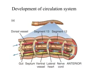
Differentiation of mesenchyme • The early heart is a simple tube which commences beating during the third week (Day 21-22). • Cardiogenic mesenchyme differentiates into three distinct cell populations: • Endocardium 心內膜 • Myocardium 心肌 • Epicardium 心外膜
Folding of the heart tube Apoptosis in the dorsal mesocardium will allow movement of the early heart tube within the pericardial cavity
The folding of the heart tube brings the inflow and outflow trunks in an adjacent position at the superior aspect of the developing heart.
The dorsal fold forms the two pericardial sinuses and places the fibrous skeleton in a single plane
Inflow and outflow trunks are positioned posteriorly as a result of the dorsal fold
Normal development of Heart • Early Events • Critical Changes • Endocardial cushion(心內墊) growth and fusion • Bulboventricular looping • Interatrial septum(心房間隔) formation • Interventricular septum(心室間隔) formation • Aortico-pulmonary septum(主肺動脈隔) formation • Vascular Changes
Endocardial cushion fusion • Endocardial cushions are areas of the fibrous skeleton forming between the atrium and ventricle. • Endocardial cushions serve two important functions: • form a partition in the heart tube between the atrium (PA) and ventricle (PV) (tricuspid and bicuspid valves) • provide a "scaffold" of the interatrial septae and the interventricular septum Defects in endocardial cushion fusion are associated with trisomies 18 and 21 (Down's syndrome).
Valvular atresia Valvular atresias arise from the uneven partition of the AV canal. Depending on the size and position of the narrowed channel, a tricuspid atresia or a biscuspid atresia results. Atresias arising from anomalous partition of the canal are referred to as congenital atresias
Bulboventricular looping BV looping is a consequence of several changes: - Dorsal folding The first dorsal fold forms an expanded primitive ventricle, referred to as the bulboventricular loop. - Ventricular growth Differential growth of the proximal ventricular tissue causes a counter-clockwise rotation of the folded heart tube. The site of ventricular growth marks the future left ventricle. Abnormal growth of the distal primitive ventricle causes clockwise rotation, an anomaly known as dextrocardia心偏右. - AV canal房室管 partitioning The Atrio-Ventricular (AV) canal between the primitive atrium and ventricle has now been partitioned by the fusing endocardial cushions. - Shunting of venous return The development of the venous system causes an increase in right-sided venous return to the primitive atrium. Combined with the partitioning of the AV canal, the change in blood flow volume and directions assists in the outgrowth of the left ventricle.
Interatrial septum formation Blue arrows - direction of growth; Red arrow - direction of blood flow;ECC - endocardial cushion; RA - right atrium; LA - left atrium.
原隔 原孔 次孔 次隔
Atrial septal defects心房間隔缺損 (ASD) Atrial septal defects (ASD) are fairly common, present in 10-15% of patients with congenital cardiac anomalies. It is more commonly observed in females than males (2-3:1).
Interventricular septum formation AP - aorticopulmonary; ECC - endocardial cushion;Blue arrow - direction of bulbar ridge growth; Red arrow - direction of ventricular growth
Aortico-pulmonary septum formation The aortico-pulmonary (AP) septum arises within the truncus arteriosus. The septum results from the downwards growth and fusion of bulbar ridges, induced by invasion of neural crest cells. The AP septum serves to divide the ventricular outflow between the pulmonary artery and the ascending aorta
Tetralogy of Fallot The tetralogy of Fallot results from the asymmetric division of the AP septum. The result is a stenosed pulmonary artery and a VSD. • Tetralogy of fallot: • Pulmonary valve stenosis • Ventricular septal defect • Overriding aorta • Hypertrophy of right ventricle
Eisenmenger's syndrome • Characteristics of Eisenmenger's syndrome: • persistent truncus arteriosus • ventricular septal defect • left-right ventricular shunt • right ventricle hypertrophy
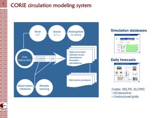
Normal development of Heart • Early Events • Critical Changes • Vascular Changes • Overview of embryonic circulatory system • Venous development • Arterial development • Anatomical correlations