Download
1 / 28
280 likes | 286 Vues
This lecture overview covers the general and special sensory receptors, anatomy and physiology of the ear and eye, as well as external anatomy of the orbital region. It includes information on the sensory organs within the head, hearing and equilibrium in the ears, sight in the eyes, smell in olfactory organs, and taste in taste buds.

E N D
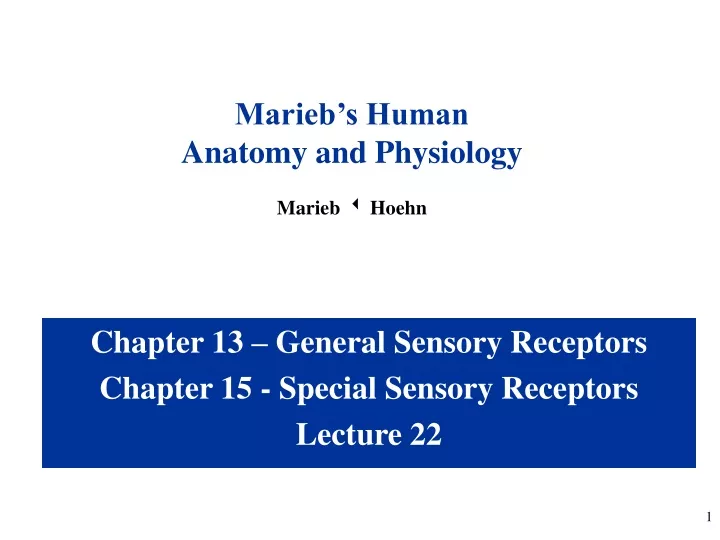
Marieb’s Human Anatomy and Physiology Marieb w Hoehn Chapter 13 – General Sensory Receptors Chapter 15 - Special Sensory Receptors Lecture 22
Lecture Overview • Introduction to the senses and sensation • Types of sensors • Classification of sensory receptors • Anatomy of the ear • Physiology of hearing/equilibrium • Anatomy of the eye • Physiology of vision Video 1 Video 2 Video 3
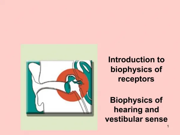
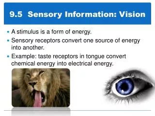
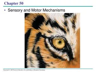
Special Senses • sensory receptors are within large, complex sensory organs in the head • hearing and equilibrium in ears • sight in eyes • smell in olfactory organs • taste (gustation) in taste buds (Video 2) (Video 3)* Not covered in video – see master slide set
External Anatomy of the Orbital Region Figure from: Saladin, Anatomy & Physiology, McGraw Hill, 2007
The Eye and Deep Orbital Region • Visual Accessory Organs • eyebrows • eyelids (palpebrae) • conjunctiva • lacrimal apparatus • extrinsic eye muscles Limbus
Eyelids • palpebrae = eyelids • composed of four layers • skin • muscle • connective tissue • conjunctiva • orbicularis oculi – closes eye (CN VII) • levator palpebrae superioris – raises eyelid (CN III) • tarsal (Meibomian) glands – secrete oil onto eyelashes; keep lids from sticking together • conjunctiva – mucous membrane; lines eyelid and covers portion of eyeball; keeps eye from drying out Fornix Sagittal section of right eye Figure from: Saladin, Anatomy & Physiology, McGraw Hill, 2007
Lacrimal (Tear) Apparatus • lacrimal gland • lateral to eye • secretes tears • canaliculi • collect tears • lacrimal sac • collects from canaliculi • nasolacrimal duct • collects from lacrimal sac • empties tears into nasal cavity Tears: - supply oxygen and nutrients to cornea (avascular) - are antibacterial (contain antibodies and lysozyme) - lubricate and bathe the conjunctiva
Extraocular Eye Muscles • Superior rectus • rotates eye up and slightly medially • Inferior rectus • rotates eye down and slightly medially • Medial rectus • rotates eye medially
Extrinsic Eye Muscles • Lateral rectus • rotates eye laterally • Superior oblique • rolls eye, rotates eye down and laterally • Inferior oblique • rolls eye, rotates eye up and laterally Which cranial nerves innervate each of the muscles in the diagram above? LR6SO4AO3
Extraocular Eye Muscles & their CN Which cranial nerves innervate each of the muscles in the diagram above? LR6SO4AO3
Structure of the Eye - Overview Figure from: Martini, Fundamentals of Anatomy & Physiology, Pearson Education, 2004 Three layers (tunics) of the eye: - Outer fibrous tunic - Sclera and cornea - Middle vascular tunic (uvea) – Iris, ciliary body, and choroid - Inner neural tunic - Retina
Outer (Fibrous) Tunic • Cornea • anterior portion • transparent • light transmission • light refraction • well innervated • avascular • Sclera • posterior portion • opaque • protection • support • attachment site for extrinsic eye muscles Transverse section, superior view
Aqueous Humor • fluid in anterior cavity of eye • secreted by epithelium on inner surface of the ciliary processes • provides nutrients • maintains shape of anterior portion of eye • leaves cavity through canal of Schlemm (scleral venous sinus)
Lens • transparent, avascular • biconvex • lies behind iris • largely composed of lens fibers • enclosed by thin elastic capsule • held in place by suspensory ligaments of ciliary body • focuses visual image on retina (Crystallins) Loss of lens transparency = cataracts
Accommodation • changing of lens shape to view objects nearby Far vision (emmetropia)(20 ft. or greater) Presbyopia is the loss of the ability to accommodate with age Near vision
Middle (Vascular) Tunic = Uvea • 1. Iris • anterior portion • pigmented CT • controls light intensity • 2. Ciliary body • anterior portion • pigmented • holds lens • muscles reshape lens for focusing • aqueous humor • 3. Choroid coat • provides blood supply • pigments absorb extra light This layer contains the intrinsic muscles of the eye- Regulate the amount of light entering the eye - Regulate the shape of the lens
Iris • composed of connective tissue and smooth muscle • pupil is hole in iris • dim light stimulates (sympathetic) radial muscles and pupil dilates • bright light stimulates (parasympathetic, CN III) circular muscles and pupil constricts mydriasis miosis How would viewing near objects affect pupil size?
Inner (Neural) Tunic • retina • containsvisual receptors • continuous with optic nerve • ends just behind margin of the ciliary body • composed of several layers • macula lutea – yellowish spot in retina surrounds fovea • fovea centralis – center of macula lutea; produces sharpest vision; only cones • optic disc – blind spot; contains no visual receptors • vitreous humor – thick gel that holds retina flat against choroid coat Visual axis Transverse section, superior view
Optic Disc (Blind Spot) Figure from: Martini, Fundamentals of Anatomy & Physiology, Benjamin Cummings, 2004
Layers of Retina • receptor cells, bipolar cells, and ganglion cells - provide pathway for impulses triggered by photoreceptors to reach the optic nerve • horizontal cells and amacrine cells – modify impulses
Visual Receptors • Rods • long, thin projections • contain light sensitive pigment calledrhodopsin • hundred times more sensitive to light than cones • provide vision in dim light • produce colorless vision • produce outlines of object • view off-center at night • Cones • short, blunt projections • contain light sensitive pigments called erythrolabe, chlorolabe, and cyanolabe (photopsins) • provide vision in bright light • produce sharp images • produce color vision Dark adaptation by the rods takes approximately 30 minutes. This adaptation can be destroyed by white light in just milliseconds
Rods and Cones Storage site of vitamin A Figure from: Martini, Fundamentals of Anatomy & Physiology, Benjamin Cummings, 2004 Retinal is chemically related to vitamin A and is made from it.
Mechanism of Light Transduction Phosphodiesterase (PDE) in the retina is a form of the enzyme that Viagra inhibits. Could this cause visual problems? Figure from: Marieb, Human Anatomy & Physiology, Pearson Education, 2004
Rods and Cones – Neural Connections Figure from: Saladin, Anatomy & Physiology, McGraw Hill, 2007 (in fovea centralis) Many rods synapse with a single bipolar cell giving poor resolution (acuity). In fovea, 1 cone synapses with one bipolar cell. Therefore, the resolution (acuity) is better using cones and they produce sharp vision.
Image Information Figure from: Martini, Fundamentals of Anatomy & Physiology, Benjamin Cummings, 2004
Stereoscopic Vision Because the pupils and fovea are 6-7 cm apart, each eye receives a slightly different image. This allows the slightly different pictures to be integrated by the brain resulting in stereoscopic vision and depth perception.
Visual Pathway The right side of the brain receives input from the left half of the visual field The left side of the brain receives input from the right half of the visual field Figure from: Martini, Fundamentals of Anatomy & Physiology, Benjamin Cummings, 2004
Please Take the Short Quiz Remember to take the brief quiz now to see if you’ve gotten the major concepts from the video. You can always go back and review the video as many times as you like – and can retake the quiz, as well.