Download
1 / 68
800 likes | 1.31k Vues
Disorders of Red Blood Cells. Professor Myat Thandar Department of Physiology University of Medicine 1. Functions of RBCs. O 2 transport (Hb in the RBCs) CO 2 transport Acid-base balance. Functional Importance of the Biconcave Shape of RBCs. Larger surface area for O 2 diffusion

E N D
Disorders of Red Blood Cells Professor Myat Thandar Department of Physiology University of Medicine 1
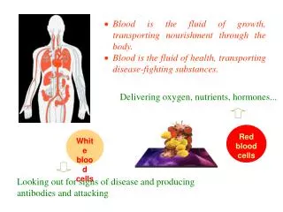
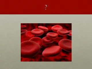
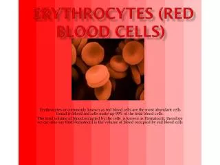
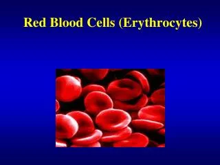
Functions of RBCs • O2 transport (Hb in the RBCs) • CO2 transport • Acid-base balance
Functional Importance of the Biconcave Shape of RBCs • Larger surface area for O2 diffusion • Thinness of cell membrane enables O2 to diffuse easily • Flexibility of membrane facilitates the transport function
Network of Fibrous Proteins of RBCs • Spectrin and Ankyrin • Imparts elasticity and stability to membrane and allows RBCs to deform easily
Haemoglobin • A natural pigment, reddish when oxygenated • 4 polypeptide chains (a globin portion and a heme unit)
Haemoglobin F in Fetus • Higher affinity for O2 than adult Hb • HbF is replaced within 6 months of birth with HbA
Haemoglobin Synthesis • Availability of iron for heme synthesis • Amount of iron: 2 g in women and 6 g in men Clinically, decreased ferritin levels usually indicate the need for prescription of iron supplements.
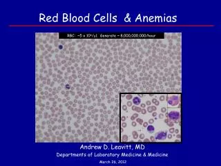
Red Cell Production • Until 5, almost all bones; After 20, membranous bones • Approximately 1% of total RBC is generated from bone marrow each day • Reticulocyte count serves as an index of erythropoietic activity of bone marrow
Stages of Erythropoiesis • Hematopoietic stem cell (HSCs) • Unipotent committed stem cell • Proerythroblast (15-20 mm) • Early normoblast (12-16 mm) • Intermediate normoblast (10-14 mm) • Haemoglobinization begins • Late normoblast (10-14 mm) • Haemoglobinization ++ • Nuclear disintegration • Reticulocyte (7-8 mm) • Haemoglobinization ++ • Nucleus remains only as strands of reticular element • Erythrocyte (7.5 mm) IL-1, IL-6, IL-3 (interleukins) GM-CSF, G-CSF, SCF Erythropoietin GM-CSF
Red Cell Maturation • Reduction in the cell size • Increase in the amount of haemoglobin • Disappearance of nucleus, and • Change in staining characteristics of cytoplasm: basophilic to eosinophilic. This is partly due to a fall in content of RNA.
Human Erythropoietin Produced by recombinant DNA technology Used for anaemia induced by chemotherapy in cancer patients, and HIV infected persons treated with zidovudine In severe anaemia, retic count may be as much as 30% (normal about 1%); numerous erythroblasts may appear in the blood
Excretion of Bilirubins Excess bilirubin elimination leads to bilirubin gallstones If red cell destruction and bilirubin production is excessive, yellow discoloration of the skin, jaundice, occurs due to accumulation of unconjugatedbilirubin
Haemoglobinuria • Haemoglobin binding protein – Haptoglobin – in the plasma • Other plasma proteins – albumin – also binds to Hb • Extensive destruction of RBCs (haemolytic transfusion reactions), binding capacity is exceeded • Haemoglobinaemia and haemoglobinuria results
Red Cell Metabolism 2,3-DPG decreases affinity of Hb for O2, facilitating the release of O2 at tissue levels Increased 2,3-DPG occurs in chronic hypoxia such as chronic lung diseases, anemia and residence at high altitude
Inhibition of Oxygen Haemoglobin Binding Certain chemicals : nitrates and sulfates Hb reacts with nitrite to form methaemoglobin G6PD deficiency predisposes to oxidative denaturation of hemoglobin with resultant red cell injury and lysis (oxidative stress generated by infection or exposure to certain drugs)
Laboratory Tests • Using automated blood cell counters: red cell content and indices • Red cell indices are used to differentiate type of anemias by size or color of red cells • Haemoglobin • Hematocrit • Mean corpuscular volume (MCV falls in microcytic and rises in macrocytic anemia) • Mean corpuscular haemoglobin concentration (normochromic or normal MCHC; hypochromic or decreased color or decreased MCHC)
Laboratory Tests • Mean cell haemoglobin • A stained blood smear: information about size, color and shape of red cells and the presence of immature or abnormal cells • If blood smear is abnormal, bone marrow examination may be indicated • Bone marrow aspiration from posterior iliac crest or the sternum
Red cell count and Haemoglobin severity of anemia Red cell characteristics Size normocytic, microcytic or macrocytic Color normochromic, hypochromic Shape the cause of anemia
Anemia • Values of hemoglobin, hematocrit or RBC counts which are more than 2 standard deviations below the mean • HGB<13.5 g/dL (men) <12 (women) • HCT<41% (men) <36 (women) Normal Hb Concentration Western value Myanmar value Male : 16 g / dL (14 - 17 g/dL) 14.4 g / dL Female : 14 g / dL (12 - 15.5 g/dL) 12.5 g / dL
Pathophysiology of Anemia • Blood Loss • Decreased Production (lack of nutritional elements or bone marrow failure) • Increased Destruction (haemolysis)
Effects of Anemia • Manifestations of impaired oxygen transport and the resultant compensatory mechanisms • Reduction in red cell indices and hemoglobin levels • Signs and symptoms associated with the pathophysiologic process that causes anemia Manifestations depend on its severity, the rapidity of its development and the person’s age and health status
Impaired oxygen transport and tissue hypoxia • Weakness, fatigue, dyspnoea and angina • Brain hypoxia results in headache, faintness and dim vision • Redistribution of blood results in pallor of skin, conjunctiva, mucous membranes and nail beds
Compensatory Mechanisms • Tachycardia, palpitations and increased cardiac output • A flow type of systolic murmur • Ventricular hypertrophy and high output heart failure • Accelerated erythropoiesis results in diffuse bone pain and sternal tenderness Haemolytic anemia : jaundice Aplastic anemia : petechiae and purpura due to reduced platelet functions
Blood Loss Anemias • Depends on rate of haemorrhage and blood loss is external or internal • Rapid loss causes circulatory shock and collapse; fall in red cell count, Hb, hematocrit due to fluid shift into vessels • Initially red cells are normocytic, normochromic • Increased erythropoietin and retic count • Slow loss (GI bleeding, menstrual disorders) causes anemia; signs and symptoms develop if the amount of red cell mass loss reach 50% (Hb <8 g/dL)(iron deficiency anemia) • External bleeding leads to iron loss and iron deficiency
HaemolyticAnemias • Characterized by premature destruction of red cells, retention in the body of iron and other products of Hb destruction and increased erythropoiesis • Normocytic normochromic red cells • Increased retic count in the circulating blood • Haemoglobinemia, haemoglobinuria, jaundice, haemosiderinuria
HaemolyticAnemias • Intravascular haemolysis is less common, caused by complement fixation in transfusion reactions, mechanical injury or toxic factors • Extravascular haemolysis occurs when RBCs are less deformable to traverse splenic sinusoids, characterized by anemia and jaundice • Intrinsic : defects of red cell membrane, haemoglobinopathies (sickle cell disease and thalassemias) and enzymes defect • Extrinsic or acquired : drugs, bacteria and other toxins, antibodies and physical trauma
Inherited Disorders of Red Cell Membrane • Hereditary spherocytosis : abnormalities of spectrin and ankyrin • Mild hemolytic anemia, jaundice, splenomegaly and bilirubin gallstones • Splenectomy done to reduce red cell destruction and blood transfusion in a crisis
Sickle Cell Disease • Haemoglobin S (point mutation in the βchain of Hb, valine for glutamic acid) • Haemolytic anaemia, jaundice, gallbladder stones, pain and organ failure (infarction of organs) • Hb S becomes sickled when deoxygenated or at a low oxygen • Deoxygenated Hb aggregates and polymerizes in the cytoplasm, creating a semisolid gel that changes the shape and deformability of the cell
Red Cell Sickling • Chronic hemolytic anemia • Blood vessel occlusion • Associated conditions: cold, stress, physical exertion, infection, illnesses that cause hypoxia, dehydration or acidosis
Diagnosis and Treatment • Neonates : clinical findings and haemoglobin electrophoresis • Prenatal diagnosis : analysis of fetal DNA by amniocentesis • Prevention of sickling episodes, symptomatic treatment and treatment of complications (prophylactic penicillin and full immunization) • Cytotoxic drug – hydroxyurea – to allow synthesis of more HbF and less HbS • Nitric oxide appears to be a promising new drug • Bone marrow or stem cell transplantation
Thalassemias • Inherited disorders of haemoglobin synthesis and decreased synthesis of αorβglobin chains of HbA • Heterozygous or homozygous
Thalassemias • βthalassemias – Cooley anaemia or Mediterranean anemia – common in Mediterranean population of southern Italy and Greece • αthalassemias more common among Asians • Anemia due to low production of affected chain and continued production and accumulation of unaffected globin chain • Reduced Hb synthesis leads to hypochromic microcytic anemia; accumulation of unaffected chain interferes with normal red cell maturation, and membrane changes leading to hemolysis and anemia
β -thalassemias • Excess α chains are denatured to form precipitates (Heinz bodies) in the bone marrow red cell precursors • Heinz bodies impair DNA synthesis and damage to red cell membrane • Coagulation abnormalities, thrombotic events (stroke and pulmonary embolism) in moderate to severe form
Pathophysiology of β -thalassemias Thinning of cortical bones
Treatment of β -thalassemias • Regular transfusion to maintain Hb at 9 to 10 g/dL • Iron chelation therapy to reduce iron load • Stem cell transplantation • Stem cell gene replacement
α-thalassemias • Synthesis of globin chains is controlled by 4 genes • Deletion of single gene : silent carrier; two genes is αthalassemia trait • Deletion of three genes leads to unstable aggregates of αchains – HbH • Four globin chains are deleted : Hb Bart (extremely high oxygen affinity, cannot release oxygen in the tissues • Chronic hemolytic anemia
Inherited Enzymes Defect • G6PD deficiency • RBCs vulnerable to oxidants, direct oxidation of Hb to methaemoglobin, and denaturation of Hb to form Heinz bodies • Anti-malaria drug primaquine, the sulfonamides, nitrofurantoin, aspirin, phenacetin, some chemotherapeutics and other drugs cause hemolysis • Diagnosed through G6PD assay or screening test
Acquired Hemolytic Anaemias • By direct membrane destruction or antibody mediated lysis • Various chemicals, toxins, venoms, malaria infection, prosthetic heart valves, vasculitis, severe burns, septicaemia, thrombotic thrombocytopenic purpura, renal disease • Warm reacting antibodies (IgG) and cold reacting antibodies (IgM) • Warm antibodies bind with Ag on red cell membrane (Rh Ag), resulting in spherocytosis and destruction by RE system • Cold antibodies activate complements; as in lymphoproliferative disorder and idiopathic
Coomb’s Test • Direct Antiglobulin Test (DAT) is positive in autoimmune hemolytic anaemia, erythroblastosis fetalis, transfusion reactions, transfusion reactions and drug induced hemolysis • Indirect antiglobulin test is used for antibody detection and crossmatching before transfusion
Anemias of Deficient Red Cell Production • Deficiency of nutrients for hemoglobin synthesis (iron) • Deficiency of nutrients for DNA synthesis (Cobalamin or folic acid) • Marrow is replaced by nonfunctional tissues
Iron Deficiency Anemia • Dietary deficiency (vegetarians) • Loss of iron through bleeding (peptic ulcer, polyps, cancer, menstrual bleeding) • Increased demands (growing children, pregnancy)