Download
1 / 53
540 likes | 1.02k Vues
Diabetic Neuropathy in the Context of Diabetic Foot. Podiatry Workshop Kuala Lumpur 18 – 19 November 2K Dr.Ashok Kumar Das Dean, Director, Prof. and Head Dept. of Medicine, JIPMER Pondicherry. Diabetic Neuropathy in the Context of Diabetic Foot - 2.

E N D
Diabetic Neuropathy in the Context of Diabetic Foot • Podiatry Workshop • Kuala Lumpur 18 – 19 November 2K • Dr.Ashok Kumar Das • Dean, Director, Prof. and Head • Dept. of Medicine, JIPMER • Pondicherry
Diabetic Neuropathy in the Context of Diabetic Foot - 2 • DFU - cause for more amputation than any • other pathology. Is it inevitable? • St. Vincent’s and Health 21 WHO Declaration • have called for reduction in amputation in • diabetic foot. • Most contemporary and challenging issue
Diabetic Neuropathy in the Context of Diabetic Foot - 3 • “5-10% of all diabetic patients have foot ulceration of various degrees and about 1% undergo amputation” • “Diabetes accounts for upto 50% of non traumatic leg amputations” • “Of all the diabetic amputees about 50% will lose their life or their other leg by 3 years”
Diabetic Neuropathy in the Context of Diabetic Foot • Foot ulcers result in • *Morbidity • * Mortality • “Enormous health care expenditure” • “Psychosocial problems” • Paucity of data regarding prevalence of • diabetic foot in India
Diabetic Neuropathy in the Context of Diabetic Foot - 5 • Neuropathy that is significant enough to cause foot ulceration may affect 40% of diabetic population especially elderly with type 2 diabetes.
Indian Patients • Our major problem is neuropathic ulcer • 85 - 95 %, • 10 – 15% vascular. • -younger patients • -mean age of amputation earlier • - number of amputations for neuropathic ulcer
Indian Context • Eminently preventable • Amputation in a diabetic neuropathic • ulcer is deplorable • Need of the hour • * Awareness/ education • * Early identification of a high risk foot • and its appropriate management
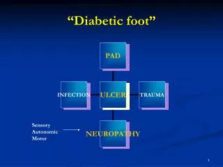
Components of neuropathic foot transforming to diabetic foot - 1 • A. Ulcerative diabetic neuropathic foot • components • B. Non-ulcerative neuropathic pathologies in diabetic foot
A. Components of ulcerative diabetic neuropathic foot - 2 • 1. Neuropathic foot and Neuroischaemic foot • 2. Neuropathic foot deformities • a. Clawed toes • b. Pes cavus • c. Hallux rigidus and valgus • d. Hammer toe • e. Nail deformities • f. Charcot foot
A. Components of ulcerative diabetic neuropathic foot - 3 • 3. Neuropathic callus • 4. Neuropathic oedema • 5. Neuropathic ulcers • a. Callus ulcers • b. Ulcer over the pressure points on the sole • c. Decubitus ulcers • d. Puncture wound ulcers • e. Traumatic ulcers
B. Non-ulcerative neuropathic pathologies in diabetic foot • 1. Charcot Foot • Acute, chronic bone destruction and • deformed diabetic foot and toes • 2. Pathological fractures • 3. Diabetic painful neuropathy
DN in context of diabetic foot - 1 • Our job – look after “NDF at risk” • A: Preventive measures • Treatment rapid and intensive • Rest • Off load • Antibiotics • Foot wear • Patient education
DN in context of diabetic foot - 2 • B. Metabolic control • Hyperglycemia • Hypertension • Hyperlipidaemia • Cessation of smoking • C. Deformity management • D. Callus management • E. Debridement and dry skin and fissure • management
DN in context of diabetic foot - 3 • F. Mechanical control • Off load, Off load, Off load • by rest, crutches, walkers, protective shoes, heel protective pad, decrease plantar pressure by removal callus • G. The importance of callus removal in NFU • decrease plantar pressure • know full dimension of the ulcer • deep swab • drainage of exudate, removal of dead tissue • H. Infection control • I. Educational control
Diabetic neuropathy Scope of the syndrome - 1 • Three components of neuropathy: • *sensory – painful, painless • *Motor • *Autonomic • All contribute to diabetic foot ulceration • Asymptomatic neuropathy 35%
Diabetic neuropathy Scope of the syndrome - 2 • Diffuse • 1. Distal symmetric sensorimotor • polyneuropathy • 2. Autonomic neuropathy • A.Sudomotor • B.Cardiovascular • C.Gastrointestinal • D.Genitourinary
Diabetic neuropathy Scope of the syndrome - 3 • Focal • 1.Cranial neuropathy • 2.Radiculopathy/plexopathy • 3.Entrapment neuropathy • 4.Asymmetric lower limb motor • neuropathy- Diabetic amyotrophy
Diabetic neuropathy Scope of the syndrome - 4 • Rather than acting in isolation neuropathy • exerts • Its vicious effects in concert with • angiopathy + immunopathy leading to • infections
Neuropathic foot components • Neuropathic ulcer • Neuropathic joint • Neuropathic oedema
Diabetic foot – Mechanisms - 1 • Loss of pain sensation results in • neuropathic injury due to • repeated unrecognised trauma • inflicted in many different ways
Diabetic foot – Mechanisms - 2 • Loss of joint positon sense results • in abnormal foot posture • This may lead to injury when • the shoes are not properly selected • or during walking
Diabetic foot – Mechanisms - 3 • Motor Neuropathy • * Weakness • * Wasting of small intrinsic muscles of • foot • *Imbalance between the flexor and extensor muscles
Diabetic foot – Mechanisms - 4 • Intrinsic deformity • Clawing of the toes • Prominence of metatarsal heads • Flattening of the arch
Diabetic foot – Mechanisms - 5 • Abnormal distribution of body weight • Weight gets concentrated on smaller • areas like metatarsal head and the heel. • Excess pressure loading of these areas • finally results in callus formation.
Diabetic foot – Mechanisms - 6 • Body weight in patients with plantar ulcers • was significantly greater than in those • with neuropathy but no ulcer.
Autonomic neuropathy - 1 • * Damages the sympathetic innervation of lower limb • * This results in • Decreased sweating • Results in dry skin fissures / cracks • Super added infection
Autonomic neuropathy - 2 • Opening of arteriovenous channels • Warm skin ( misleadingly healthy ) • Shunting of nutrients and oxygen from • the tissues • Impaired vascular response to infection
Autonomic neuropathyclassical signs • Dry skin with fissuring • Distended veins over the dorsum of foot • and the ankle
Connective tissue changes • Hyperglycemia causes non enzymatic • glycation of collagen and keratin • Increase in cross linking • Become rigid and inflexible • Tissue break down in places where there • is high horizontal shear force
Neuropathic ulcer • * Painless • * Develops on pressure points (metatarsal heads/heel) * Pulsations intact unless superadded ischaemia is also present * Decrease in pain / temperature as also in the • vibration perception • * Punched out ulcer surrounded by callus
Neuropathic (n) /Ischemic ulcer (i) • Site Pressure points (n) • Sides / tips of toes (i) • Pain --- ( n ) +++ ( i ) • Callus ++ ( n ) --- ( i ) • Pulse ++ ( n ) --- ( i ) • Abi > 1 ( n ) < .6 ( i ) • Healing ++ ( n ) --- ( i )
Quantitative tests for neuropathic assessement - 1 • 1. Measurement of light touch sensation • - Nylon monofilament (Semmes • Weinstein) • 2. Measurement of thermal sensitivity • - Marstock Thermode
Quantitative tests for neuropathic assessement - 2 • The advantage of the assessment with monofilaments or biosthesiometry is the detection of whether the patient has lost protective pain sensation that would render him susceptible to foot ulceration. • Nylon monofilaments test the threshold to pressure sensation
Quantitative tests for neuropathic assessement - 3 • Monofilament: This is a simple technique. • When applied perpendicular to the foot it • buckles at a force of 10 gms • Areas to be tested include plantar aspect • of big toe metatarsal heads of first, third • and fifth and the plantar surface of heel. • Filament not to be applied over the callus
Quantitative tests forneuropathic assessement - 4 • 1. Measurement of vibration • *Biosthesiometer • *Graduated tuning fork • 2. Nerve conduction studies
Management of Neuropathic Ulcer - 1 • General measures • Specific measures
Management of Neuropathic Ulcer - 2 • Good glycemic control • Treatment of infections • Management of neuropathic oedema
Management of Neuropathic Ulcer - 3 • All ulcers irrespective of their cause will be slow to heal in presence of oedema, due impairment of local flow • Neuropathic oedema can be treated with • Diuretics • Ace inhibitors • Ephedrine ( 30 mg tds ) • D/d hypo albuminemia cardiac failure
Management of Neuropathic Ulcer - 4 • “Over 90% of predominantly neuropathic • ulcers will heal satisfactorily with • conservative measures”
Management of Neuropathic Ulcer - 5 • “Key to the management is the relief of • pressure that caused the initial lesion” • Pressure is off loaded most effectively by • encasing the foot in a light plaster of paris • cast. • *Total contact cast • *Removable scotch cast boot, custom made shoes etc.
Preventing Neuropathic Foot Ulcers - 1 • Regular inspection of foot - annually • Identification of high risk feet – 3 mo / 6mo • Careful choice of foot wear • Regular chiropody • Intense education
Preventing neuropathic foot ulcers - 2 • “As little as one hour’s education provided by the podiatrist resulted in 70% reduction in amputations over the following 2 years . as compared with a control group who did not receive the advice” • Malone IM et al 1989
Glycemic control and diabetic neuropathy • Diabetes control and complication trial • showed that intensive insulin therapy • reduced the incidence of appearance of • neuropathy by about 70%
Intensive insulin treatment • Reduced the clinical appearance of overt • neuropathy in patients with subclinical • neuropathy from 16% to 7% (57% • reduction) • Reduced the risk of developing clinically • overt diabetic neuropathy by 60% over five • years
Neuropathic joint or Charcot arthropathy - 1 • 1868 French neurologist I.M. Charcot • First described in tabes • Can also be seen in leprosy, • syringomyelia, • hereditary sensory neuropathy, • Charcot Marie Tooth disease etc
Neuropathic joint or Charcot arthropathy - 2 • Relatively rare • Potentially devastating disorder • Long standing diabetes • Dense peripheral neuropathy • Peripheral vascular disease is typically • absent
Neuropathic joint or Charcot arthropathy - 1 • Sympathetic failure-- increased blood • flow due to arteriovenous anastomosis • Bone demineralisation (diabetic • osteopenia) • Susceptibility to minor, recurrent fractures
Neuropathic joint or Charcot arthropathy - 4 • Painless disintegration of bone in • response to trivial trauma • Common joints involved are • Tarso metatarsal • Metatarso phalangeal • Ankle joint • Knee joint
Neuropathic joint or Charcot arthropathy - 5 • Acute Charcot arthropathy may mimic infection • Chronic Charcot foot is classically described as ‘bag of bones’ • (Gross destruction of joint surfaces and bone with effusion which is typically painless)
Neuropathic joint or Charcot arthropathy - 6 • Differentiation from osteomyelitis is • difficult • * TC 99 Scan • * Indium labelled white cell scan • * MRI