Download
1 / 1
20 likes | 132 Vues
A hand viewed through a mirror on the prototype sonic flashlight. Development of a Hybrid Ultrasound Biopsy Phantom to Study The Effect of Tissue Compressibility on Target Identification and Localization Gaurav Shukla , Bing Wu PhD, Roberty Klatzky PhD, Jules Sumkin DO, George Stetten MD,PhD

E N D
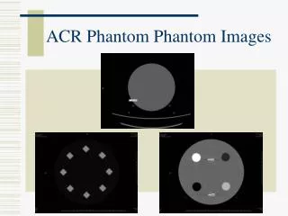
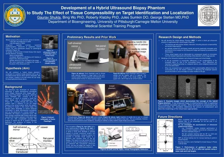
A hand viewed through a mirror on the prototype sonic flashlight Development of a Hybrid Ultrasound Biopsy Phantom to Study The Effect of Tissue Compressibility on Target Identification and Localization Gaurav Shukla, Bing Wu PhD, Roberty Klatzky PhD, Jules Sumkin DO, George Stetten MD,PhD Department of Bioengineering, University of Pittsburgh/Carnegie Mellon University Medical Scientist Training Program Motivation Preliminary Results and Prior Work Research Design and Methods • Breast cancer is the most common cancer in women. • >200,000 new diagnoses in 2006 • >40,000 deaths in 2006 • Diagnosis typically involves screening (mammography), ultrasound of suspicious lesions (Figure 1a), and subsequent biopsy for pathological evaluation (Figure 1b). • A number of issues arise in breast biopsy that require special attention, including: • tissue compressibility • increased difficulty in identifying and keeping a target in the ultrasound image • greater reliance on the fine detail in the image • the extra requirement of keeping the needle in the plane of the ultrasound scan • We will develop the Hybrid Biopsy Phantom (HBP), in which a blank gel ultrasound phantom is augmented with virtual targets (Figure 6) • The physical gel will provide realistic interaction including physical compressibility and actual image speckle • An optically tracked SF and biopsy needle will permit graphically simulated cysts and tumors to be overlaid on the ultrasound image and targeted at predetermined 3D locations • These virtual targets will be deformable by compression based on information from the tracked implements and tracking of the distortion of the image speckle • Modeling of the interaction between real ultrasound images and virtual targets • It will be necessary to incorporate differences in the compressibility of the simulated lesion vs. that of the surrounding tissue. Such differences are important in diagnosing benign vs. malignant lesions, since malignant lesions tend to be less deformable., • When the simulated lesion is modeled to have a deformability different from the surrounding gel, we will explore methods of deforming the displayed speckle around the lesion to accommodate its motion relative to the gel. Figure 1a: ultrasound image of a fluid-filled cyst in breast tissue. Figure 1b: ultrasound image of cyst aspiration (needle visible) Hypothesis (Aim) We will construct a “hybrid biopsy phantom,” comprised of a realistic breast phantom and a set of simulated breast lesions which will be integrated with one another, creating a tool with which we can study the psychophysical issues above. Figure 3a (above): Sonic flashlight used in clinical trials placing catheters in deep veins of the arm. Custom plastic housing attaches half-silvered mirror and flat-panel monitor to ultrasound transducer. Figure 3b (above): Sonic flashlight used to guide a needle into the jugular vein of a cadaver. The needle tip is visible in the virtual image of the jugular vein, which floats at its correct location. Background Our laboratory has developed an innovative technology to guide interventional procedures which merges ultrasound (US) images with a direct view of the patient. The device, called the Sonic Flashlight (SF), consists of a US transducer, on which is mounted a small flat-panel display and a semitransparent mirror (Figure 2). Looking through the mirror, the operator sees a reflection of the US image floating at the actual location within the patient. By aiming for targets in this virtual image, the operator takes advantage of natural hand-eye coordination to guide percutaneous procedures such as the placement of a venous catheter, or potentially, the biopsy a suspected tumor. We have recently conducted NIH-funded clinical trials using the SF to place catheters in the deep veins of the arm, with considerable success. We propose to extend the SF to another important application: the guidance of biopsy in the diagnosis of breast cancer. Figure 6: Example images which demonstrate the concept of the hybrid biopsy phantom. A real ultrasound image of a blank phantom (left) is augmented with a computer-simulated cyst (center). When the phantom is compressed by the ultrasound probe, the simulation determines the amount of compression and alters the morphology of the virtual lesion appropriately. Future Directions Figure 2 (below): schematic of Sonic Flashlight. Through-plane (Figure 4a, above, left) and in-plane (Figure 4b, above, right) insertion of needle into a cyst in a breast phantom (BluePhantomTM). In-plane insertion (right) shows the shaft of the needle in the scan. For our purposes, however, these physical phantoms have limitations, including an inability to easily redefine target shape and location. • When completed, the HBP will demonstrate a number of features that, to our knowledge, do not yet exist in an ultrasound phantom • We will investigate the psychophysics of ultrasound guided breast biopsy: • e.g., Comparison studies between performance in guidance tasks between conventional ultrasound and the sonic flashlight (Figure 7) • We will compare learning with the SF vs. CUS and develop training procedures based on the HBP. • HBP as clinical training tool for novices learning to perform breast biopsies • Develop and test a sonic flashlight for breast biopsy in Phase I clinical trials The conception (Figure 5a, near right) and a prior implementation (Figure 5b, far right) of a virtual tomographic reflection (VTR) system, in which a mock sonic flashlight is optically tracked to provide continuous location and orientation information. Using the tracker, it is possible to simulate virtual targets in a phantom and visualize them on the SF display. Prior work in our lab utilized this VTR system to study the psychophysics of the SF as compared with conventional ultrasound (CUS), and we will adapt this system in the development of the hybrid biopsy phantom. Figure 7: Performance of guidance tasks using conventional US vs. SF in a fully-real system using a water tank and real targets Figures 5a (left) and 5b (right): Virtual Tomographic Reflection