Download
1 / 59
600 likes | 813 Vues
Advances in Bioscience Education Summer Workshop . Fluorescence and Electron Microscopy June 26 - 29, 2007 Biological Electron Microscope Facility Pacific Biosciences Research Center University of Hawai’i at Manoa. What is a Microscope?.

E N D
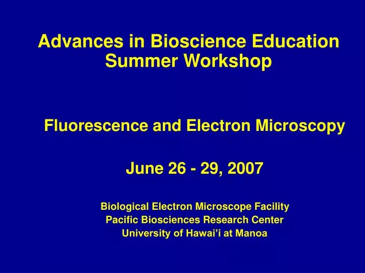
Advances in Bioscience Education Summer Workshop Fluorescence and Electron Microscopy June 26 - 29, 2007 Biological Electron Microscope Facility Pacific Biosciences Research Center University of Hawai’i at Manoa
What is a Microscope? • A tool that magnifies and improves resolution of the components of a structure • Has three components: • sources of illumination, • a magnifying system, • detectors.
Sources of Illumination • Light microscopes use a beam of light for illumination and include fluorescence and confocal microscopes • Electron microscopes use electrons as a source of illumination and include transmission and scanning electron microscopes.
Lenses are used to control a beam of illumination, magnify, and direct an image to a detector Light and Electron Microscopes
Common FluorescenceApplications • Localize/identify specific organelles • Detect live cells vs. dead cells, necrotic vs. apoptotic cells • Determine cell membrane permeability • Localize antigen-specific molecules • Multiple labeling
Laser Scanning Confocal Microscope • Better resolution • Serial optical sections can be collected from thick specimens • Live or fixed cell and tissue imaging
Laser Scanning Confocal Microscopy Drosophila eye Plant Protoplast Photos courtesy of Gregg Meada & Dr. Gert DeCouet, UHM And Dr. Chris Yuen and Dr. David Christopher
Epifluorescence vs. Confocal Sample courtesy Gregg Meada & Dr. Gert DeCouet, UHM
Scanning Electron Microscopy (SEM) • View outer surface • Coat specimen with gold • No sectioning • High Mag (40x to 300,000x) • High resolution (better than 2 nm)
Transmission Electron Microscopy(TEM) View inside cell via sections magnification 120,000 X 50,000X
Conventional TEM Micrographs Bacteria in cell Apoptosis Skin Collagen Virus in cell Chloroplast
Ultra-microtomy • Ultrathin (60-90 nm) sectioning of resin-embedded specimens • Several brands/models available
Cryotechniques • Ultrarapid cryofixation • Metal mirror impact • Liquid propane plunge • Freeze fracture with Balzers 400T • Cryosubstitution • Cryoultramicrotomy – Ultrathin frozen sections (primarily for antibody labeling)
Immunolocalization • LM • Fluor/confocal • TEM • SEM with backscatter detector
Approaches to Immunolabeling • Direct Method: Primary antibody contains label • Indirect Method: Primary antibody followed by labeled secondary antibody • Amplified Method: Methods to add more reporter to labeled site
Two-step Indirect Method for Immunolabeling • Fluorescent-conjugated secondary antibody attaches to primary antibody that is bound to antigen
Immunolabeling for Transmission Electron Microscopy • Normally do Two-Step Method • Primary antibody applied followed by colloidal gold-labeled secondary antibody • May also be enhanced with silver
Colloidal Gold Immunolabeling for TEM • Colloidal gold of defined sizes, e.g., 5 nm, 10 nm, 20 nm, easily conjugated to antibodies • Results in small, round, electron-dense label easily detected with EM • Can be enhanced after labeling to enlarge size for LM or EM
Double-labeling Method • Use primary antibodies derived from different animals (e.g., one mouse antibody and one rabbit antibody) • Then use two different secondary antibodies conjugated with different sized gold particles
Preparation of Biological Specimens for Immunolabeling • Preserve tissue as closely as possible to its natural state while at the same time maintaining the ability of the antigen to react with the antibody • Chemical fixation OR • Cryofixation
Chemical Fixation • Antigenic sites are easily denatured or masked during chemical fixation • Glutaraldehyde gives good fixation but may mask antigens, plus it is fluorescent • Paraformaldehyde often better choice, but results in poor morphology , especially for electron microscopy • May use e.g., 4% paraformaldehyde with 0.5% glutaraldehyde as a good compromise
Embedding • Dehydrated tissue is embedded in a plastic resin to make it easier to cut thin sections
Steps in Labeling of Sections • Chemical fixation • Dehydration, infiltration, embedding and sectioning • Blocking • Incubation with primary antibody • Washing • Incubation with secondary antibody congugated with reporter (fluorescent probe, colloidal gold) • Washing, optional counterstaining • Mount and view
Controls! Controls! Controls! • Omit primary antibody • Irrelevant primary antibody • Pre-immune serum • Perform positive control • Check for autofluorescence • Check for non-specific labeling • Dilution series
Light Path in Fluorescence • Light delivered through excitation filter and then objective lens to specimen where it is absorbed; • emitted light goes back through objective lens through barrier filter and emission filter and then to detector.
Fluorescence • Light beam excites the fluorochrome, raising it to a higher energy state, • As it falls back to it’s original state, it releases energy in the form of a light of lower E and longer wavelength than original beam of light
Primary Ab = PDIsecondary Ab = AlexafluorBlue light = exciting beamgreen and red light emitted • And use them to your advantage! • Green is label; orange-red is autofluorescence • Acts as counterstain Know Your ArtifactsAutofluorescence
Fluorescence • Fluorochromes are excited by specific wavelengths of light and emit specific wavelengths of a lower energy (longer wavelength)
Filter Cubes for Fluorescence • Filter cubes generally have an excitation filter, a dichroic element, and an emission filter • The elements of a cube are selected for the excitation and fluorescence detection desired
Laser Scanning Confocal Microscopy • Fluorescence technique • Uses laser light for excitation • Improves image resolution over conventional fluorescence techniques • Optically removes out-of-focus light and detects only signal from focal plane • Can construct an in-focus image of considerable depth from a stack of images taken from different focal planes of a thick specimen • Can then make a 3-D image that can be tilted, rotated, and sliced
Principal Light Pathway in Confocal Microscopy • Laser light is scanned pixel by pixel across the sample through the objective lens • Fluorescent light is reflected back through the objective and filters (dichroic mirrors) • Adjustable pinhole apertures for PMTs eliminate out-of-focus flare • Image is detected by photomultiplier(s) and digitized on computer
TEM • Transmission Electron Microscope • Illumination source is beam of electrons from tungsten wire • Electromagnetic lenses perform same function as glass lenses in LM • Higher resolution and higher magnification of thin specimens
Specimen Preparation for TEM • Chemical fixation with buffered glutaraldehyde • Or 4% paraformaldehyde with >1% glutaraldehyde • Postfixation with osmium tetroxide • Or not, or with subsequent removal from sections • Dehydration and infiltration with liquid epoxy or acrylic resin • Polymerization of hard blocks by heat or UV • Ultramicrotomy – 60-80nm sections • Labeling and/or staining • View with TEM
High pressure freezing: Plant tissue is flash frozen in a pressure bomb -197 C Water in the tissue is replaced with acetone over 5 day period Acetone saturated tissue is embedded in resin Resin is cut in thin sections, 80 nm thick Add antibodies - immunolabeling Look under Electron microscope
Very Wrinkled
Chloroplast Carnage Pretty bad fixation
2nd time: stainings were done poorly, but there is hope… Back to the drawing board to start over. But what to correct? What to do different? Will it improve?
Despite mistakes, keep moving forward and ignore doubt and negativism that comes with pressure.
3rd time A charm
Excellent preservation And Immunolabeling the 3rd TIME
HIGH MAG
RE-search Not search Must be repeated Research time is spent: 70% trouble-shooting 15% success 15% communicating success.