Download
1 / 44
440 likes | 449 Vues
Biochemistry-I Spring Semester 2015. Course Outline Chapters as in the text Unit I: Protein Structure and Function 1: Amino acids 2: Structure of Proteins 3: Globular Proteins 4: Fibrous Proteins 5: Enzymes 6: Chemistry of lipids 7: Introduction to Carbohydrates 8: Glycolysis

E N D
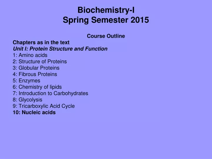
Biochemistry-ISpring Semester 2015 Course Outline Chapters as in the text Unit I: Protein Structure and Function 1: Amino acids 2: Structure of Proteins 3: Globular Proteins 4: Fibrous Proteins 5: Enzymes 6: Chemistry of lipids 7: Introduction to Carbohydrates 8: Glycolysis 9: Tricarboxylic Acid Cycle 10: Nucleic acids
Textbook: . Biochemistry, 3rd edition. Harvey and Champe (editors). Champe, Harvey, and Ferrier. Lippincott Williams & Wilkins (2005). • StryerBiochemistry: A short course, Tymoczko J.l., Berg J.M. and Stryer L., W.H. freeman and Company , New York, (2010). • Lenhinger Principles of Biochemistry, David L. Nelson and Michael M. Cox, 4th edition, W.H. freeman and Company (2004). • Midterm exam 30% • Quizes 20% • Final Exam 50% Contact details • Prof. Dr. B. M. ZABUT • Biochemistry & Nutrition • bzabut@iugaza.edu
Amino Acids UNIT I: Protein Structure and Function
Overview • Proteins: most abundant and functionally diverse • Examples: • Enzymes and polypeptide hormones direct and regulate metabolism • Contractile proteins movement • Collagen + calcium phosphate crystals bone • Hemoglobin and plasma albumin in blood shuttle molecules • Immunoglobulins fight infectious agents • All are polymers of amino acids
Structure of the amino acids • More than 300 have been described in nature • Only 20 (encoded by DNA) are commonly found in mammalian proteins • Each (except proline) has a carboxyl group, an amino group, and a distinctive side chain (R-group)
At physiologic pH (~ 7.4), -COOH COO- and NH2 NH3+ • In proteins, these groups are involved in peptide linkage not available for chemical except for H-bonding • Side chain dictates role of aa in a protein • aa’s classified according to side chains
A. Amino acids with non-polar side chains • R-group does not bind or give off protons or participate in hydrogen or ionic bonds • R-groups can be thought of as “oily” or “lipid like” a property that promotes hydrophobic interactions.
Non-polar amino acids • Location in proteins: • In proteins found in aqueous solutions, side chains tend to cluster in interior of protein i.e., fill up interior of folded protein help give 3D shape. • In proteins located in membranes, non-polar R-groups are on outside surface, interacting with lipid env.
2. Proline • The side chain and α-amino group form a ring • Thus it contains imino group, rather than amino • Its unique geometry contributes to formation of fibrous structure of collagen, and often interrupts α-helices found in globular proteins
B. Amino acids with uncharged polar side chains • Zero net charge at neutral pH • Side chains of cysteine and tyrosine can lose protons at alkaline pH • Serine, threonine, tyrosine hydroxyl groups can participate in H-bonding • R-groups of asparagine and glutamine, containing a carbonyl and amide group each, can also participate in H-bonding
Disulfide bond • R-group of Cys contains –SH group, an important component of active site of many enzymes • In proteins –SH groups of two cysteines can become oxidized to form a dimer, cystine, which contains a covalent crosslink called a disulfide bond (-S-S-)
2. Side chains as sites of attachment for other compounds • Ser, Thr, and rarely Tyr, contain a polar hydroxyl group as site of attachment for structures e.g., phosphate group. • Amide group of Asn, as well as hydroxyl group of Ser or Thr, can be sites of attachment for oligosaccharide chains in glycoproteins
C. Amino acids with acidic side chains • Amino acids Asp and Glu are proton donors • At neutral pH, R-groups fully ionized, containing a negatively charged carboxylate group (–COO-) • Therefore called aspartate and glutamate to emphasize that they are negatively charged at physiologic pH
C. Amino acids with basic side chains • R-groups of basic aa’s accept protons • At physiologic pH, R-groups of Lys, and Arg are fully ionized and positively charged • Histidine is weakly basic, free aa is largely uncharged at physiologic pH. When in protein, His R-group can be either positive or neutral depending on the ionic env. provided by the polypeptide chains of the protein. • This contributes to role of His in functioning of proteins such as Hb
E. Abbreviations and symbols for the commonly occurring amino acids • Three-letter abbreviation and one-letter symbols • The one-letter codes are determined by following rules: • Unique first letter: if only one aa begins with a particular letter e.g., I = ileucine • Most commonly occurring aa’s have priority e.g., gly is more common than glutamate, so G = glycine • Similar sounding names: e.g., F = phenylalanine, W = tryptophan • Letter close to initial letter: for remaining aa’s symbol is the letter as close in alphabet as possible to the initial of the aa e.g., K = lysine. • Further, B is assigned to Asx, Z to Glx, and X to unidentified aa.
Figure 1.7. Abbreviations and symbols for the commonly occurring amino acids.
F. Optical properties of amino acids • The α-carbon of each aa is attached to 4 different chemical groups i.e., a chiral or optically active carbon atom • Gly is an exception why? It is optically inactive • aa’s that have an asymmetric center at the α-carbon can exist in two forms D and L • The two forms in each pair termed stereoisomers, optical isomers, or enantiomers • All aa’s found in proteins are of the L-form • D-aa’s are found in some antibiotics and in bacterial CW
III. Acidic and Basic Properties of amino acids • aa’s in aqueous solution contain weakly acidic α-carbonyl and weakly basic groups α-amino groups • Additionally, acidic and basic aa’s contain ionizable groups • Thus, free aa’s and some aa’s in peptide linkages can act as buffers • The quantitative relation b/w the conc. of weak acid (HA) and weak base (A-) is described by Henderson-Hasselbalch equation
A. Derivation of the equation • HA H+ + A- • Ka = [H+] [A-] [HA] • The larger the Ka, the stronger the acid and vice versa • By solving the above equation, you obtain the Henderson-Hasselbalch equation • pH = pKa + log [A-] [HA]
B. Buffers • Resist change in pH following addition of an acid or base • Created by mixing weak acid (HA) with its conjugate base (A-) • If acid (e.g., HCl) is added, A- can neutralize it, being converted into HA • If base is added, HA can neutralize it, being converted into A- • Maximum buffering capacity occurs at pH = pKa, but still HA/A- pair can serve as an effective buffer when pH is within ~ ± 1 pH unit of the pKa
C. Titration of an amino acid • Dissociation of the carboxyl group: • As for other weak acids • e.g., Ala with an α-carboxyl and an α-amino group: at a low (acidic) pH, both groups are protonated • As pH raised, -COOH group of form I can dissociate by donating a proton to medium COO- (form II, a dipolar form) this form is a.k.a zwitterion, the isoelectric form of Ala
Figure 1.10. Ionic forms of alanine in acidic, neutral, and basic solutions.
2. Application of the Henderson-Hasselbalch eq. • Dissociation constant of carboxyl group is called K1 • The Henderson-Hasselbalch eq. K1 = [H+] [II] [I] pH = pK1+ log [II] [I]
3. Dissociation of the amino group: • The second titratable group of Ala is -NH3+ • A much weaker acid than the –COOH, therefore a much smaller dissociation constant K2 (i.e., pK2 is larger) • Release of a proton from -NH3+ fully deprotonated form of Ala (form III)
4. pKs of alanine • Each titratable group has a pKa that is numerically equal to the pH at which exactly one half of the protons have been removed from that group
5. Titration curve of alanine Figure 1.11. The titration curve of alanine.
Note the following: • Buffer pairs: the –COOH/-COO- pair can serve as a buffer in the pH region around pK1, and the –NH3/ -NH2 pair can buffer in the region around pK2 • When pH=pK: when pH = pK1(2.3), equal amounts of forms I and II exist in solution. When pH = pK2 (9.1), equal amounts of form II and form III. • Isoelectric point: at neutral pH, alanine exists as the dipolar form II, net charge is zero. Isoelectric point (pI) is the pH at which aa is electrically neutral. For aa that has only two dissociable hydrogens, pI = (pK1 + pK2)/2, i.e., (2.3 + 9.1)/2 = 5.7. pI corresponds to pH at which structure II predominates, and at which equal amounts of form I (net charge of +1) and III (net -1) exist
6. Net charge of amino acids at neutral pH • At physiologic pH, all aa’s have a negative –COO- and a positive –NH3+ group, both attached to the α-carbon • Glu, Asp, His, Arg, Lys have additional potentially charged groups • Substances, e.g., aa’s, that can act as an acid and a base are defined as amphoteric, and are referred to as ampholytes (amphoteric electrolytes)
D. Other applications of Henderson-Hasselbalch eq. • Can be used to calculate how the pH of a physiologic solution responds to changes in the conc. weak acid and/or its corresponding “salt” form. • E.g., in bicarbonate buffersystem, the equation predicts how shifts in [HCO3-] and pCO2 influence pH
Figure 1.12 A. changes in pH as the concentrations of HCO3- or CO2 are altered;
The eq. is also useful for calculating the abundance of ionic forms of acidic and basic drugs. E.g., most drugs are either weak acids or weak bases. • Acidic drugs (HA): HA ↔ H+ + A- • Weak bases (BH+) can also release a H+. The protonated form of a basic drug is usually charged, and loss of a H+ uncharged base (B): BH+ ↔ B + H+
A drug passes through membranes more readily if uncharged. • For a weak acid, the uncharged (HA) can permeate through membranes and A- cannot. • For a weak base, such as morphine, the uncharged, B, penetrates and BH+ does not
The effective conc. of permeable form at its absorption site is determined by relative conc. of charged/uncharged forms. • The ratio is determined by pH at site of absorption, and by strength of the weak acid or base which is represented by pKa of the ionizable group • The eq. is useful in determining how much drug found on either side of a membrane separating two compartments that differ in pH e.g., stomach (pH 1.0-1.5) and blood plasma (pH 7.4)
Summary • Each aa has an α-carboxyl and an α-amino group (except Pro, has an imino group) • At physiologic pH: α-carboxyl COO-, α-amino NH3+ • Each aa contains one of 20 side chains • The chemical nature of side chain determines function of aa in a protein, and classifies aa as nonpolar, uncharged polar, acidic, or basic • All free aa’s plus charged aa’s in peptide chains serve as buffers • Relation b/w the conc. of weak acid and its conjugate base is described by Henderson-Hasselbalch eq. • Buffering occurs within ±1 pH, and max when pH = pKa at which [HA] = [A-] • The α-carbon of each aa (except Gly) is attached to 4 different chemical groups i.e., a chiral or optically active carbon atom • Only L-form of aa’s is found in proteins synthesized by human body