Download
1 / 72
720 likes | 727 Vues
This module covers topics such as neuromuscular junction events, G proteins and pathways, alkalosis and acidosis, genetics, endocrinology, mitosis/meiosis, cell cycle, and oncogenes/tumour suppressor genes.

E N D
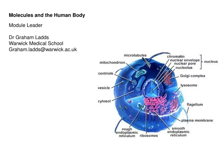
Molecules and the Human Body Module Leader Dr Graham Ladds Warwick Medical School Graham.ladds@warwick.ac.uk
Signalling • Neuromuscular junction events • G proteins – pathways • Alkalosis and acidosis • Genetics – group work in lecture theater • Preparation for next few weeks • The ‘bolics’ • Endocrinology • Mitosis/meiosis • Cell cycle • Oncogenes/tumour suppressor genes • ESA style questions Topics to cover today
For once size does matter Size Name Units Example 10-3 10-6 10-9 10-12 10-15 10-18 milli micro nano pico femto atto m µ n p f a mM µL ng pM fL ag Drug levels Concentration A 1M solution = 1 mole/Litre A 1mM solution = 10-3 mole/Litre moles = mass(g) e.g. 1 mole of carbon = 12g MW 12 1 mole = 6.022x10²³ molecules – Avogadro’s number
Signal > Translator > Response • The translator detects the signal - it is a receptor. • The translator converts the signal into the response - it is an effector. • The translator is a protein (or series of proteins). • Effectors use three mechanisms to change cell behaviour. • 1. Alter gene transcription. • 2. Alter ion balance across the plasma membrane. • Alter the activity level of existing enzymes. • There are five types of receptor • 1. Intracellular receptors. • 2. Receptors that are ion channels. • 3. Receptors with intrinsic enzyme activity. • 4. Receptors linked to protein kinases. • 5. Receptors coupled to target proteins via a G protein.
Alter gene transcription Changing the protein composition changes cell behaviour. Some genes are turned on. Some genes are turned off. Not suitable for rapid, short-term changes. Common mechanism for development and differentiation.
Alter ion balance across the plasma membrane Changing ion balance changes cell behaviour. Transport of Na+, K+ or Cl- changes membrane potential. Transport of Ca2+ changes intracellular concentration. Ca2+ is a second messenger (affects activity of target proteins).
Many signals alter the activity level of enzymes Many enzymes affect protein phosphorylation. Protein kinases (phosphorylate target proteins). Protein phosphatases (dephosphorylate target proteins). Many enzymes affect second messenger levels. Phospholipase C (hydrolyses PIP2 to IP3 and DAG). Adenylate cyclase (converts ATP to cAMP). Guanylate cyclase (converts GTP to cGMP). cGMP phosphodiesterase (converts cGMP to GMP). Phosphoinositide 3-kinase (phosphorylates phosphoinositides).
C=O C=O C=O C=O O O CH CH CH2 P OH P OH 6 5 P OH HO NH 2 P OH 1 4 N IP3 N OH P OH N N 2 3 Ca2+ O CH 2 O PtdIns(3)P 4 1 3’5’-cyclic AMP 3 2 O P O OH O Second messengers Concentration of second messenger changes after stimulation. Second messengers regulate the activity of target proteins. Exoplasm O O Cytoplasm CH2 CH CH2OH Diacylglycerol
Signal > Translator > Response • The translator detects the signal - it is a receptor. • The translator converts the signal into the response - it is an effector. • The translator is a protein (or series of proteins). • Effectors use three mechanisms to change cell behaviour. • 1. Alter gene transcription. • 2. Alter ion balance across the plasma membrane. • Alter the activity level of existing enzymes. • There are five types of receptor • 1. Intracellular receptors. • 2. Receptors that are ion channels. • 3. Receptors with intrinsic enzyme activity. • 4. Receptors linked to protein kinases. • 5. Receptors coupled to target proteins via a G protein.
Intracellular receptors that are enzymes Activating receptor changes their enzyme activity. Some enzymes become more active. Some enzymes become less active. Changing enzyme activity changes cell behaviour. Nitric oxide and intracellular guanylate cyclase. NO* diffuses across the membrane and binds to guanylate cyclase. Guanylate cyclase converts GTP to cGMP (a second messenger). cGMP affects the activity of target proteins (protein kinase G). NO* is used in many signalling pathways. Controls blood vessel dilation (amyl nitrate spray). Allows peristaltic movement through the gut.
Signal > Translator > Response • The translator detects the signal - it is a receptor. • The translator converts the signal into the response - it is an effector. • The translator is a protein (or series of proteins). • Effectors use three mechanisms to change cell behaviour. • 1. Alter gene transcription. • 2. Alter ion balance across the plasma membrane. • Alter the activity level of existing enzymes. • There are five types of receptor • 1. Intracellular receptors. • 2. Receptors that are ion channels. • 3. Receptors with intrinsic enzyme activity. • 4. Receptors linked to protein kinases. • 5. Receptors coupled to target proteins via a G protein.
The opening of ion channels Voltage-gated channels (changes in membrane potential). Channels for Na+, K+ and Ca2+. Ligand-gated channels (extracellular ligands - neurotransmitters). Excitatory transmitters open Na+/K+-channels (depolarisation). Acetylcholine (nicotinic receptor) – sympathetic NS . Glutamate. Serotonin (5HT-3 receptor).
The 6 events that occur when a nerve impulse reaches the NMJ resulting in neurotransmitter release 1.Voltage-regulated calcium channels in the axon membrane open. 2. Allows Ca2+ to enter the axon. 3. Ca2+ inside the axon terminal causes some of the synaptic vesicles to fuse with the axon membrane. 4. Release of acetylcholine into the synaptic cleft (exocytosis). 5. acetylcholine diffuses across the synaptic cleft and attaches to acetylcholine receptors on the sarcolemma. 6. Binding of acetylcholine to receptors on the sarcolemma initiates an action potential in the muscle.
The opening of ion channels Voltage-gated channels (changes in membrane potential). Channels for Na+, K+ and Ca2+. Ligand-gated channels (extracellular ligands - neurotransmitters). Excitatory transmitters open Na+/K+-channels (depolarisation). Acetylcholine (nicotinic receptor) – sympathetic NS . Glutamate. Serotonin (5HT-3 receptor). Inhibitory transmitters open Cl- channels (hyperpolarisation). g-aminobutyric acid (GABA). Glycine.
The opening of ion channels Voltage-gated channels (changes in membrane potential). Channels for Na+, K+ and Ca2+. Ligand-gated channels (extracellular ligands - neurotransmitters). Excitatory transmitters open Na+/K+-channels (depolarisation). Acetylcholine (nicotinic receptor) – sympathetic NS . Glutamate. Serotonin (5HT-3 receptor). Inhibitory transmitters open Cl- channels (hyperpolarisation). g-aminobutyric acid (GABA). Glycine. Ligand-gated channels (intracellular ligands - second messengers). cAMP (olfaction), cGMP (phototransduction), Ca2+.
The opening of ion channels Voltage-gated channels (changes in membrane potential). Channels for Na+, K+ and Ca2+. Ligand-gated channels (extracellular ligands - neurotransmitters). Excitatory transmitters open Na+/K+-channels (depolarisation). Acetylcholine (nicotinic receptor) – sympathetic NS . Glutamate. Serotonin (5HT-3 receptor). Inhibitory transmitters open Cl- channels (hyperpolarisation). g-aminobutyric acid (GABA). Glycine. Ligand-gated channels (intracellular ligands - second messengers). cAMP (olfaction), cGMP (phototransduction), Ca2+. Mechanically-gated channels (sound, touch, stretch).
Signal > Translator > Response • The translator detects the signal - it is a receptor. • The translator converts the signal into the response - it is an effector. • The translator is a protein (or series of proteins). • Effectors use three mechanisms to change cell behaviour. • 1. Alter gene transcription. • 2. Alter ion balance across the plasma membrane. • Alter the activity level of existing enzymes. • There are five types of receptor • 1. Intracellular receptors. • 2. Receptors that are ion channels. • 3. Receptors with intrinsic enzyme activity. – cancer stuff. • 4. Receptors linked to protein kinases. • 5. Receptors coupled to target proteins via a G protein.
Signal > Translator > Response • The translator detects the signal - it is a receptor. • The translator converts the signal into the response - it is an effector. • The translator is a protein (or series of proteins). • Effectors use three mechanisms to change cell behaviour. • 1. Alter gene transcription. • 2. Alter ion balance across the plasma membrane. • Alter the activity level of existing enzymes. • There are five types of receptor • 1. Intracellular receptors. • 2. Receptors that are ion channels. • 3. Receptors with intrinsic enzyme activity. • 4. Receptors linked to protein kinases. – EPO. • 5. Receptors coupled to target proteins via a G protein.
Signal > Translator > Response • The translator detects the signal - it is a receptor. • The translator converts the signal into the response - it is an effector. • The translator is a protein (or series of proteins). • Effectors use three mechanisms to change cell behaviour. • 1. Alter gene transcription. • 2. Alter ion balance across the plasma membrane. • Alter the activity level of existing enzymes. • There are five types of receptor • 1. Intracellular receptors. • 2. Receptors that are ion channels. • 3. Receptors with intrinsic enzyme activity. • 4. Receptors linked to protein kinases. – EPO. • 5. Receptors coupled to target proteins via a G protein.
Ligand G protein-coupled receptors (GPCRs) g g g g a a a a and b b b b b b b b GTP GDP GTP GDP Nucleotide exchange Activate target proteins Transcription factors Ion channels Protein kinases and phosphatases Phospholipase C Phosphoinositide 3-kinase Cyclases and phosphodiesterases GTP GDP
Variations on a very common theme Human cells are estimated to have at least 1000 GPCRs. Neurotransmitters, hormones, lipids, chemokines, odours. Human cells contain many different types of G proteins. There are at least 20 Ga subunits. There are at least 5 Gb subunits. There are at least 12 Gg subunits. Some ligands bind to more than one GPCR. Some GPCRs activate more than one G protein. Dissociated subunits can regulate more than one target protein. Some target proteins are regulated by more than one G protein.
If you really want a simple version Gas stimulates adenylate cyclase. Glucagon, ACTH. Gai inhibits adenylate cyclase. Prostaglandin PGE1, adenosine. Gat stimulates cGMP phosphodiesterase. Photons (rhodopsin). Gaq stimulates phospholipase C. Bombesin, vasopressin. Ga13 activates ion channels (Na+/H+ exchange). Thrombin.
Dopamine There are D1-like and D2-like receptors. D1 and D5 couple through Gs to stimulate adenylate cyclase. D2, D3 and D4 couple through Gi to inhibit adenylate cyclase. Acetylcholine – parasymathetic ns There are five muscarinic acetylcholine receptor subtypes. M1, M3 and M5 couple through Gq to stimulate phospholipase C. M2 couples through Gi to open a K+-channel. M4 couples through Gi to inhibit adenylate cyclase. ….and don’t forget the nicotinic acetylcholine receptor. This is a Na+/K+-channel.
Serotonin There are 15 serotonin receptor subtypes. 5HT-1 couple through Gi to inhibit adenylate cyclase. 5HT-2 couple through Gq to stimulate phospholipase C. 5HT-4, 5, 6 and 7 couple through Gs to stimulate adenylate cyclase. ….and don’t forget the 5HT-3 receptor. This is a Na+/K+-channel. 5HT = 5-hydroxytryptamine. Adrenergic receptors Multiple receptors for adrenaline (epinephrine) and noradrenaline. a1 receptors couple through Gq to stimulate phospholipase C. a2 receptors couple through Gi to inhibit adenylate cyclase. b receptors couple through Gs to stimulate adenylate cyclase.
Signal integration in cardiomyocytes Contraction is regulated by stimulatory and inhibitory signals. b-adrenergic receptors stimulate adenylate cyclase. a-adrenergic receptors inhibit adenylate cyclase Both receptors work through G proteins. Adenylate cyclase converts ATP to cAMP (a second messenger). b-adrenergic receptor a-adrenergic receptor Stimulate Inhibit Adenylate cyclase Gi protein Gs protein cAMP ATP
Signalling • Neuromuscular junction events • G proteins – pathways • Alkalosis and acidosis • Genetics – group work in lecture theater • Preparation for next few weeks • The ‘bolics’ • Endocrinology • Mitosis/meiosis • Cell cycle • Oncogenes/tumour suppressor genes • ESA style questions Topics to cover today
100 pH 7.4 80 pH 7.2 60 Saturation (%) 40 pO2 in muscle capillaries 20 0 0 10 20 30 40 50 60 70 80 90 100 pO2 (torr) The Bohr effect Metabolically active tissues generate H+. The pH of the blood can be reduced from 7.4 to 7.2. The presence of H+ lowers the O2 affinity of Hb (O2 is released). Hb release ~10% more O2 at pH 7.2 than pH 7.4. This helps to deliver more O2 to active muscles.
Molecular explanation of the Bohr effect The protons (H+) bind to particular residues in Hb. N-terminalamino group of the a-chains, Hisa122 and Hisb146. Positively charged groups form new electrostatic bonds. For example, Hisb146 interacts with Aspb94. The additional interactions stabilise deoxy-Hb (the T-form). Deoxy-Hb has a lower O2 affinity than oxy-Hb. Thus, protonation reduces the O2 affinity of Hb.
CO2 lowers the O2 affinity of Hb CO2 is converted (by carbonic anhydrase) to HCO3- and H+. H+ reduces the O2 affinity of Hb via the Bohr effect (as before). Some HCO3- reacts with N-terminal amino groups of Hb chains. Carbamation makes these groups negatively charged. These form electrostatic bonds with positive groups in Hb. The additional interactions stabilise deoxy-Hb (T-form). Thus, CO2 reduces the O2 affinity of Hb. H O- + H+ N C N+H3 + HCO3- Hb Hb Carbamino-Hb O
CO2+H2O H2CO3 HCO3- + H+ COO + 2H+ NH3 + CO2 N H Transport of CO2 from the tissuesto the Lungs • 70-80% is transported back to the lungs dissolved in the blood as bicarbonate (HCO3-) • 20-30% is transported back to lungs attached to Hb Erythrocyte Carbonic anhydrase
Conditions in the lungs promote addition of O2 to Hb and release of CO2 1. Hb(H) + O2 HbO2 + H+ 2. Hb(H) + CO2 HbCO2 3. CO2+H2O H2CO3 HCO3- + H+ Lowering [Hb(H)] forces eqn 2 to the left promoting unloading of CO2 from carbaminoHb Increasing [H+] forces eqn 3 to the left forcing CO2 out of solution
6.8 Acid-base Balance • Normal blood pH is from 7.35 - 7.45 • Going outside of this range can be very dangerous - even deadly. acidosis pH 7.0 7.35 7.45 alkalosis 7.8
Acid-base Balance • pH balance is maintained by buffers in the blood. The primary buffers are: • Bicarbonate • CO2 + H2O H2CO3 HCO3- + H+ • Phosphate • H2PO4- HPO42- + H+ • Plasma proteins - various
The carbonate system contributes the most to Blood pH control • HH eqn • pH (of blood) = pKa + Log [A-] (= base = HCO3) • [HA] (= acid = CO2) • the pKa of the CO2/HCO3 system is 6.1 • Normal pCO2 blood = 40mmHg = 1.2mM (0.03 Ksol) • Normal HCO3 blood = 24mM • therefore Log 24mM = 1.3 + 6.1 = 7.40 1.2mM
Respiratory Control of Blood pH • Blood pH can be maintained by controlling the level of CO2 exhaled. CO2 + H2O H2CO3 HCO3- + H+ • Hyperventilation • Increase in rate and depth of breathing. • Reduces the CO2 in blood, increasing pH. • Hypoventilation • Reduced rate and depth of breathing. • Increases CO2 in blood, decreasing pH.
Respiratory Acidosis and Alkalosis • When breathing patterns are improper, the following can occur. • Respiratory alkalosis (exam stress?) • Caused by hyperventilation. • Treatment - rebreathe air or administer CO2. • Respiratory acidosis • Caused by inadequate breathing. • Treatment - administer HCO3- via IV.
Metabolic Acidosis and Alkalosis • Various metabolic conditions can also create improper pH levels in the blood. • Metabolic acidosis • Can be caused by uncontrolled diabetes mellitus, diarrhea, aspirin overdose and after heavy exercise. • Note that hyperventilation, while a cause of respiratory alkalosis, can be a response to metabolic acidosis. • Metabolic alkalosis • Caused by prolonged vomiting, excessive use of bicarbonate for treating an upset stomach.
Allosteric regulation of Hb - a summary O2 affinity is determined by electrostatic interactions at a-b interface. Deoxy-Hb has more interactions and lower O2 affinity. Oxy-Hb has fewer interactions and higher O2 affinity. O2 increases the O2 affinity of Hb (cooperative binding). Structural changes reduce interactions at the a-b interface. H+, CO2 and BPG lower the O2 affinity of Hb. They all increase electrostatic interactions at the a-b interface. Protonation (H+) creates electrostatic bonds between chains. Carbamation (CO2) creates electrostatic bonds between chains. BPG forms electrostatic interactions with the polypeptide chains.
Signalling • Neuromuscular junction events • G proteins – pathways • Alkalosis and acidosis • Genetics – group work in lecture theater • Preparation for next few weeks • The ‘bolics’ • Endocrinology • Mitosis/meiosis • Cell cycle • Oncogenes/tumour suppressor genes • ESA style questions Topics to cover today
The ‘bolics (processes) • Ana-bolic • To build up • (think anabolic steroids-these are the sex steroids) • The synthesis of muscle proteins • The synthesis of adipose tissue • Storage of glucose • Cata-bolic • To break down • The breakdown of muscle • The breakdown of adipose • Breakdown of glycogen
Carbohydrate ‘terms’/process • Glycolysis • The lysis of glucose (usage thereof) • Gluco(se)neogenesis • The synthesis of new glucose • Glycogenolysis • The lysis of glycogen • Glycogen synthesis • The synthesis of glycogen
Fat/protein ‘terms’/processes • Lipolysis • Breakdown of fats • Lipogenesis • Synthesis of fats • Proteolysis • Breakdown of proteins • Protein synthesis • synthesis of proteins We cover little on this please look up yourself
Major organ/systems involved • Liver • Brain • Muscle • Stomach/GI tract • Pancreas • Hypothalamus Pituitary Adrenals (HPA) axis • Cf Repro HPO-vary axis