Download
1 / 46
520 likes | 939 Vues
Echocardiography of Cardiac Amyloidosis. Frederick L. Ruberg, MD Boston University Medical Center May 25, 2005. What is amyloid. Any misfolded protein that aggregates as a -sheet stains with Congo Red (birefringence) Implication in pathogensis of alzheimers disease ( amyloid)

E N D
Echocardiography of Cardiac Amyloidosis Frederick L. Ruberg, MD Boston University Medical Center May 25, 2005
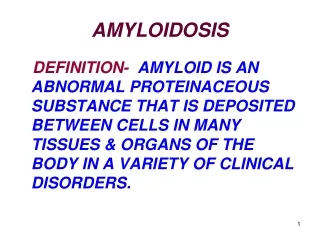
What is amyloid • Any misfolded protein that aggregates as a -sheet • stains with Congo Red (birefringence) • Implication in pathogensis of alzheimers disease ( amyloid) • Systemic amyloidoses
The Systemic Amyloidoses • Primary (AL) or light chain disease • Plasma cell dyscrasia (clonal proliferation) • 12-15% patients with myeloma have AL • Immunoglobulin light chains • 12 month survival without treatment • 6 month survival with cardiac disease • Incidence is 1 in 100,000 in Western countries • Familial (AF) • Mutations in transthyretin (TTR) • Ile 122 of particular interest
The Systemic Amyloidoses • Senile systemic amyloid (SSA) • TTR-based non-genetic (ie, TTR normal) • Cardiac predilection • Male gender, onset after age 60 • Secondary amyloidosis (AA) • Chronic inflammatory states • Other specific protein abnormalities • apolipoprotein A-I and A-II, lysozyme
Manifestations of AL Merlini, G. et al. N Engl J Med 2003;349:583-596
Diagnosis of Amyloidosis Falk, R. H. et al. N Engl J Med 1997
Amyloid Cardiomyopathy • Very poor prognosis (6 mo survival) • Restrictive cardiomyopathy with profound abnormalities of diastolic function • Systolic dysfunction late manifestation • Classic teaching • biventricular thickening in a small ventricle • valvular thickening, “speckled pattern” • Atrial enlargement • Pericardial effusion/evidence of elevated filling pressures
Echo Features Rehman, JACC 2004
Amyloid Cardiomyopathy • Patients do NOT respond to normal medication for CHF • ACE inhibitors, beta-blockers, dig • There is a treatment for AL amyloid • Autologous bone marrow transplant • Patient selection critical • assessment of cardiac involvement
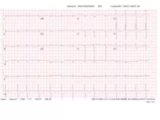
Continuum of Amyloid • Advanced disease is too late • Initial changes are abnormalities of diastolic function • As wall thickness progresses restrictive physiology ensues • Loss of limb lead voltage on ECG • Systolic dysfunction late stage
Diastolic dysfunction • Transmitral inflow • E and A wave pattern • E wave deceleration time • IVRT • Tissue Doppler mitral annular velocities • E prime < 6 cm/s • LA enlargement, IVC dilation • Restrictive physiology a late manifestation
Atrial arrest • Absent A wave in setting of NSR • Restrictive pattern • Atrial amyloid infiltration and/or markedly elevated LV DP • Risk of stroke/TIA, anticoagulation • Recovery of A wave following successful BMT correlating to symptomatic improvement
Treatment of AL • Autonomic dysfunction, low stroke volumes • Dependent on HR • Beta blockers, ACEI poorly tolerated • Digoxin may bind to amyloid and promote toxicity • Can use diuretics • Loop diuretics • Aldactone/eplerenone • Amiodarone • Proamatine (Midodrine) for BP support
SSA Clinical Features • Onset age greater than 60 years • Often exclusively cardiomyopathy • More benign clinical course than AL • Often tolerate medications that AL patients won’t • TTR amyloid, must exclude AL as well as known mutations in TTR to diagnose
Familial Amyloid CMP • Over 80 mutations identified • Ile 122 in African Americans • 2-4% heterozygotic allele frequency • Unclear penetrance • Unclear importance in setting of HTN • Onset of CMP after age 60 years • Stabilization of TTR tetramer to stop amyloidogensis by diflunisal • Other agents in development • Liver transplant/heart transplant
Stem Cell Transplant • AL can respond to chemotherapy • High dose melphalan with autologous stem cell transplantation • 8-year follow-up data (Skinner, et al. Ann Int Med 2004) • Median survival 1.6 yrs • Exclusion EF < 40% or decompensated CHF • Lower dose, marrow sparing regimens • Oral therapy, investigative drug regimens
Survival after HDM/SCT Skinner, et al. Ann Int Med 2004
Post BMT • Symptomatic improvement without obvious change in echo appearance • Hemodynamic recovery (A wave) • Improvement in TDI • BNP normalization • Mass regression • Chamber remodeling
Role of CMR • More sensitive than echo • Explore tissue-dependent changes through delayed enhancement • Demonstrated in 70% patients (Maceira, Pennell, et al. Circ 2005) associated with mass • Small LV size + increased wall thickness does not necessarily = increased mass
New echo approaches • Strain imaging determines impaired longitudinal contraction (Koyama, Falk, et. al. Circ 2003) • In absence of fractional shortening abnormality • Preceded CHF symptoms • Utility of TDI with BNP to facilitate diagnosis in early disease
Applications of echo/CMR • Early diagnosis • Predict outcomes with treatment • Monitor response to treatment