Download
1 / 65
650 likes | 799 Vues
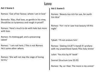
Tainted Love. Introduction. Function: Transport materials around body Components: Heart Blood Vessels. The Heart. Layers in Cross Section: Pericardium - outermost sac enclosing heart Pericardial Fluid- fluid between pericardium and epicardium

E N D
Introduction • Function: • Transport materials around body • Components: • Heart • Blood Vessels
The Heart • Layers in Cross Section: • Pericardium- outermost sac enclosing heart • Pericardial Fluid- fluid between pericardium and epicardium • Epicardium- tight fitting layer surrounding heart; also called visceral pericardium • Myocardium- cardiac muscle layer • Endocardium- smooth inner layer of heart
Heart Structure • Four chambers: • Right and left atria- receive blood into heart • Right and left ventricle- pump blood back out of the heart • Two sides are separated by septum
Valves • Four Valves in Heart: • Tricuspid - between right atrium and right ventricle • Pulmonary Semilunar- between right ventricle and pulmonary trunk • Mitral (Bicuspid) - between left atrium and left ventricle • Aortic semilunar- between left ventricle and aorta
Two Circulations of Blood • Pulmonary: • Back and forth to lungs • Systemic: • Back and forth to body
Exit Slip • 1) What chamber is this? • 2) Which valve is between right atrium and right ventricle? • 3) Which circuit (pulmonary or systemic) brings blood back and forth to lungs? • 1) Right atrium • 2) Tricuspid • 3) Pulmonary
Vessels Supplying the Heart • Coronary arteries • First two branches off of the aorta • Supply blood to heart • Cardiac veins • Return blood from heart tissues • Drain into coronary sinus • Coronary sinus • Returns blood back to right atrium
Cardiac Cycle • Sequence of events that occur during every regular heartbeat • Systole - contraction • Diastole - relaxation • Refer to timeline Systole/Diastole Song
Heart Sounds • Lubb - sound of atrioventricular (AV) valves closing • Dupp - sound of semilunar valves closing
Reminder about Cardiac Tissue • Complex network of interconnecting cells • Connected by intercalated discs • Allows them to transfer impulse rapidly and work together (functional syncytium) • Two sets in heart: • One in atria, one in ventricles • Kept separate from each other
Cardiac Conduction Intro • Electrical impulses cause heart structures to contract • Travel down a system of specialized fibers
Pathway for Conduction • Sinoatrial node (SA node) • Pacemaker • Causes atria to contract • Junctional Fibers • Delay impulse reaching ventricle by their small diameter • Atrioventricular node (AV node) • Purkinje fibers • Cause ventricles to contract
Electrocardiogram • Also know as ECG • Electrical recording of myocardium during cardiac cycle • P wave • Atrial depolarization • QRS complex • Ventricle depolarization and atrialrepolarization • T wave • Ventricle repolarization
Control of Heart Rate • Cardiac Center of Medulla Oblongata • Parasympathetic • Constant braking action; acetylcholine • Sympathetic • Increases heart rate; norepinephrine • Blood Pressure Receptors • Decreases heart rate • Impulses from Cerebrum and Hypothalamus • Decrease heart rate • Changes in K and Ca concentrations
Thumbs Up, Down • Coronary arteries supply blood to heart. • UP! • The lubb of your heart is the sound of the AV closing/opening. • UP! • An ECG measures your blood pressure. • DOWN! It measures your cardiac cycle.
Blood Vessels • System of closed tubes filled with blood • Arteries • Carry blood away from heart • Arterioles • Smaller branches of arteries • Capillaries • Thin-walled vessels where nutrients, fluid, gases, and wastes are exchanged • Venules • Small veins • Veins • Large vessels returning blood to heart
Layers of Blood Vessel Walls • Tunica externa • Outermost layer composed of connective tissue with some elastic and collagenous fibers • Tunica media • Middle layer composed of smooth muscle and elastic fibers • Tunica interna (endothelium) • Single layer of squamous epithelium
Control of Vessel Diameter • Vasoconstriction • Sympathetic nervous system impulses cause vessels to constrict • Vasodialation • Inhibition of impulse causes dialation
Arteries • Carry blood away from heart under high pressure • Has the thickest tunica media and tunica externa of all blood vessels
Arterioles • Smaller branches of arteries • Walls thin as the vessels get smaller • Eventually lose tunic externa
Capillaries • Site of exchange • Only tunica interna remains • Has small openings between endothelial cells where materials can leak out • Pre-capillary sphincters • Smooth muscle at start of capillary that can close the capillary bed and divert blood flow
Exchange of Materials • Oxygen and nutrients diffuse out of the capillary • Carbon dioxide and wastes diffuse back into capillary • Plasma Proteins don’t leave the blood • Fluid is forced out of the capillary at the arteriole side due to blood pressure • Fluid is brought back into the capillary due to osmotic pressure at the venule side • Fluid not recollected is brought back to the blood through the lymphatic system