Download
1 / 7
70 likes | 237 Vues
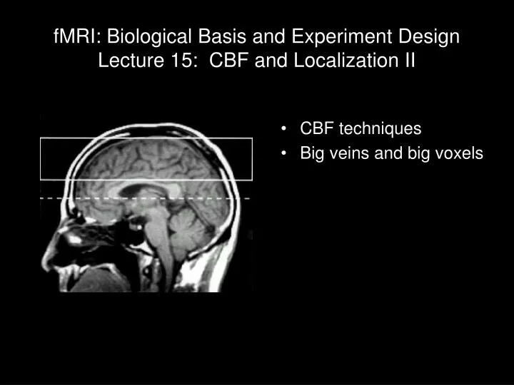
fMRI: Biological Basis and Experiment Design Lecture 15: CBF and Localization II. CBF techniques Big veins and big voxels. 1 light year = 5,913,000,000,000 miles?. Harrison, Harel et al., Cerebral Cortex 12:225 (2002). 100 m. Perfusion techniques.

E N D
fMRI: Biological Basis and Experiment DesignLecture 15: CBF and Localization II • CBF techniques • Big veins and big voxels 1 light year = 5,913,000,000,000 miles?
Perfusion techniques • FAIR – Flow-sensitive Alternating Inverstion Recovery • MOTIVE – MOdulation of TIssue and VEssel signal • QUIPSS – QUantitative Imaging of Perfusion using a Single Subtraction • QUIPSS-II – QUIPSS ... 2nd version • CASL – Continuous Arterial Spin Labeling • PASL – Pulsed Arterial Spin Labeling
Perfusion (ASL) – general idea • Water in blood is used as a tracer • Two images are measured • With inversion of incoming blood • Longitudinal magnetization of incoming blood is inverted, and subtracts from total voxel signal • Without inversion of incoming blood • Difference image indicates
CASL vs. PASL • Continuous ASL • long pulse is applied to neck arteries, so all blood coming into the head is inverted • After an appropriate delay (~1 – 2s) for that blood to get to the volume of interest, image is acquired • Reference image is taken without inversion pulse • Subtraction provides perfusion map • Pulsed ASL • Inversion is done in slab surrounding slices of interest • The rest is the same ...
Low res SE High res SE