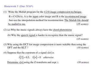
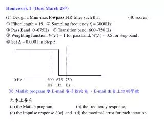
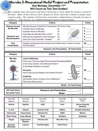
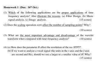
Download
1 / 9
90 likes | 175 Vues
Exams due 9am 16 th . (grades due 10am 19 th ) Describe the organization of visual signals in extra-striate visual cortex and the specialization of cells in these areas . Include a discussion of dorsal and ventral streams.

E N D
Exams due 9am 16th. (grades due 10am 19th) Describe the organization of visual signals in extra-striate visual cortex and the specialization of cells in these areas. Include a discussion of dorsal and ventral streams. 2. Describe the somatosensory system, including peripheral sensors and central projections. How does it resemble/differ from the visual system? 3. Describe the organization of the motor system. Describe the specific role of the different structures, including the evidence for that role. 4. What are Central Pattern Generators? Describe the evidence for CPG’s. What behaviors do they control? 1. Describe the roles of the basal ganglia (including reward circuitry) and the cerebellum in control of movement. 2. Why do we move our eyes? Describe the different kinds of eye movements and their properties, and the underlying neural circuitry. 3. What is meant by attention? How is it limited? Describe the evidence for a fronto-parietal attention network. 4. What is the role of the prefrontal cortex? Describe evidence for this role, including anatomical projections, fMRI, lesions, and neural recordings.
Describe the organization of visual signals in extra-striate visual cortex and the specialization of cells in these areas. Include a discussion of dorsal and ventral streams. Lecture 1 Slides 26-50 Lecture 2 slides 3-14, 22-30 Describe areas and their specialization – V2 V3 Ventral: V4, TE TEO Dorsal MT, MST LIP 2. Describe the somatosensory system, including peripheral sensors and central projections. How does it resemble/differ from the visual system? Proximal vs distal Peripheral receptive fields: X and Y cells trade off spatial and temporal sensitivity versus 4 types in somat. Sys Overall faster RT and higher temporal resolution Spatial resolution varies with receptor density Sensory quality depends on pattern of activity across receptors Somatotopicvsretinotopic – cortical magnification of fovea and hand – slide 32 Cortex – somatosensory – parallel plus serial projections slide 21 – vision mostly serial Columnar org in S! Cf V1 RF’s somewhat similar (Slide 28 and get larger at higher levels of processing – slide 30 Ventral and dorsal streams – slides 22 Active versus passive touch – slide 33 – harder to compare
More complex analysis of image properties in higher visual areas (extra-striate) Defining visual areas: Retinotopic responses Anatomical projections (cell properties) Note old simplistic view: One area, one attribute Is not true. Areas are selective in complex and poorly understood ways Note the case of Mike May.
Describe the organization of the motor system. Describe the specific role of the different structures, including the evidence for that role. Cortical areas – posterior pariteal, pre-mootr, supplementary motor, M1, basal ganglia, cerebellum, brain stem, spinal cord and motor neurons. Lecture 5 slide 4-7 Lecture 6 slides 4-14 18-47 4. What are Central Pattern Generators? Describe the evidence for CPG’s. What behaviors do they control? Stepping, swimming etc CPG’s lect 5 slide 27-31 . Describe the roles of the basal ganglia (including reward circuitry) and the cerebellum in control of movement. Lecture 7 slides 2-18 Why do we move our eyes? Describe the different kinds of eye movements and their properties, and the underlying neural circuitry. Lec 7 Slide 21-66
. What is meant by attention? How is it limited? Describe the evidence for a fronto-parietal attention network. Lecture 11 slide 10 and following Selection of stimulus or action – also spotlight vs Bayesian approach, search templates Neural – biased competition, signal ampitude, noise reduction, changes in coherence of beta waves Limits? Change blindness, dual-task interference Space, object based attention Fronto-parietal – fMRI expts, single unit evidence for attentional effects in LIP, FEF. FEF effects on V4 What is the role of the prefrontal cortex? Describe evidence for this role, including anatomical projections, fMRI, lesions, and neural recordings. Cognitive control – L 12 S 6, 26 – summarize projections S7 Specific effects of lesions – human: affective response (GSR) and input to decisions (gambling tests), impulsivity (egStroop), Action Disorganization Syndrome (Tower of Hanoi, Wisconsin card sorting) Lesions – monkey – spatial and object memory tasksk fMRI – S 10 Stroop, working memory rewards Monkey physiol – task effects on neural firing memory, rule-based activity, conditional visuo-motor associations dopamine modulation, reward effects
Attention is a hypothetical internal variable with limited/no explanatory power. Central idea: selection – of stimulus and action Also: resource limitations – not necessarily a problem, but an inevitable aspect of goal directed behavior. Ie it wouldn’t help if we were able to process more information – however, learning allows more compact codes. Potential role of basal ganglia:
What is attention? • Capacity to select information from the environment and select actions to • perform • Substantial overlap between circuitry for eye movements and circuitry for • spatial attention. • Parietal – frontal network influences visual cortical areas including V1. • LGN may gate incoming visual signals. • Attention appears to act in a way that biases competition between stimuli • within a receptive field. • Attention is limited - why? • Limitations may derive from multiple levels of processing in the brain • eg sensory, motor, and sub-cortical circuitry such as basal ganglia.
What does the cerebellum have to do with the maintenance of attention as described in the section about ADHD? If these deficits are only present in tasks with limited interest or heightened critical resources why is the cerebellum implicated? Can activity in the prefrontal cortex be used to differentiate between goal-directed and habitual behaviors? And, if a goal-directed behavior transitions to a habitual behavior will it have a different pattern of neural activation?